

 PDF
PDF ePub
ePub Citation
 Print
Print


INTRODUCTION
Epidemiological studies have reported that patients with type 2 diabetes mellitus (DM) have increased mortality and morbidity from cardiovascular diseases, independent of other risk factors (1, 2). Therefore, early detection of risk factors related to atherosclerosis is necessary to decrease these risk in patients with type 2 DM. A number of methods have been applied for assessment of cardiovascular risk and subclinical atherosclerosis. Especially, non-invasive modalities such as flow-mediated dilatation (FMD) of brachial artery, pulse wave velocity (PWV) and carotid intima-media thickness (IMT) have been widely used in clinical settings, because they have been shown to be good surrogate markers of atherosclerosis (3-5).
Arterial stiffness has been shown to be an independent predictor of cardiovascular mortality and become a useful index in the prevention and early detection of cardiovascular disease. PWV reflects arterial stiffness, and it is a marker of both the severity of vascular damage and the prognosis of cardiovascular diseases in patients with hypertension or with diabetes (6-8). Therefore, PWV is currently considered optimal, as recently described in the consensus statement on arterial stiffness (9). Recently, brachial-ankle pulse wave velocity (baPWV) using a volume rendering method is simple, noninvasive method which correlates well with arterial stiffness. And it is a useful tool for identifying a subgroup in the population that are at increased risk for cardiovascular events (10).
Many factors such as age, smoking, dyslipidemia, hypertension, microalbuminuria, waist hip ratio, HbA1c, γGTP, C-reactive protein, duration of diabetes and metabolic syndrome are the known factors to affect arterial stiffness (11-17). So far, these factors affecting baPWV have been reported in various population such as patients with type 2 diabetes, hypertensive patients and metabolic syndrome. However, determinants of baPWV in normotensive young adults with type 2 diabetes have not been examined yet. This population is relatively in early stage of DM, and there is less concern about diabetic complications. So, to evaluate which factors are related to arterial stiffness in this population can be meaningful. Herein, the purpose of this study was to assess major factors affecting baPWV in normotensive young adults with type 2 DM.
MATERIALS AND METHODS
Patients
We measured baPWV with a noninvasive pulse wave analyzer (form PWV/ABI, Colin Co., Komaki, Japan) in 2,728 consecutive type 2 diabetic patients in Department of Endocrinology and Metabolism, Pusan National University Hospital, between January 2008 to January 2011. Among these, we retrospectively included patients aged between 30 and 39 yr (n = 145). Those who had hypertension, current antihypertensive medication, atherosclerotic vascular disease, chronic kidney disease (eGFR < 60 mL/min/1.73 m2), moderate to severe heart disease (previous coronary artery disease and heart failure with NYHA class II-IV) and low ankle brachial index (< 0.9) were excluded. Presence of hypertension was defined when the subject was taking anti-hypertensive medication or measured systolic or diastolic blood pressures were same or above 130/80 mmHg. A total of 103 patients with type 2 DM were finally included in this study.
Laboratory assessment
Blood pressures (BP) were taken with a standardized sphygmomanometer after at least 5 min of rest. The height and weight of the subjects were measured just before examining baPWV. The body mass index (BMI) was expressed as the weight in kilograms divided by squared height in meters (kg/m2). The waist circumference was measured in the standing position, at the level of umbilicus by a single examiner. Blood samples were collected after at least 8 hr of fasting. Plasma glucose, serum insulin, C-peptide, lipid profile, high sensitive-C-reactive protein (hs-CRP), aspartate aminotransferase (AST), alanine aminotransferase (ALT), gamma glutamyl transpeptidase (γ-GTP) were checked concurrently in study day. HbA1c was measured by high performance liquid chromatography using the Variant TM II Turbo (Bio-Rad Laboratories, Hercules, CA, USA). Also, we investigated history of smoking, alcohol intake and medication by questionnaire.
Assessment of arterial stiffness
After the subject had rested in a supine position for more than 5 min, the measurement of baPWV was conducted using a waveform analyzer (VP-2000; Colin Co Ltd, Komaki, Japan). Electrocardiographic electrodes were placed on both wrists, a microphone was placed on the left edge of the sternum to detect heart sounds, and pneumatic cuffs were placed on both arms and ankles. The cuffs were connected to a sensor that determined volume pulse form and an oscillometric pressure sensor that measured blood pressure. The characteristic points of waveforms were determined automatically according to the pulse velocity theory. The components over 5 Hz were stored using a pass filter and the wave front was determined. The time interval between the wave front of brachial waveform and that of ankle waveform was defined as the time interval between brachium and ankle (ΔTba). The distance between sampling points of baPWV was calculated automatically according to the subject's height. The path length from the heart to the brachium (Lb) was measured externally and was expressed using the following equation: Lb = 0.2195 × height of the patient (cm) - 2.0734. The path length from the heart to the ankle (La) was expressed using the following equation: La = 0.8129 × height of the patient (cm) + 12.328. Finally, baPWV was calculated from the following equation: baPWV = (La-Lb)/ΔTba (18).
Statistical analysis
The statistical analysis was performed using the SPSS (SPSS ver 17.0 for Windows, Chicago, IL, USA) software package. All data were expressed by mean ± S.D. for normally distributed values and median (interquartile range) for nonparametric values. Pearson chi-square test was used to assess differences in categorical data between male and female. And Student t-test and Mann Whitney U test were used to assess numerical data as appropriate. Bivariate correlation (Pearson's correlation coefficient) analyses were performed to analyze the relationships between maximal baPWV and other numerical data. To approximate normal distribution, natural log transformed value for Urine Albumin/Creatinine ratio (microalbuminuria) was used in this analysis. Linear regression analysis was performed to evaluate the association between maximal baPWV and other clinical data. And step-wise multiple regression analysis was performed to determine the correlation and independent variables for maximal baPWV. A value of P < 0.05 was considered to be statistically significant.
RESULTS
Clinical characteristics of the patients
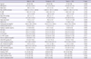
Table 1 shows the general characteristics of the study population. Pulse, fasting glucose, γ-GTP, hs-CRP, serum creatinine, smoking, alcohol intake and statin treatment were different significantly between male and female.
Correlations between maximal baPWV and other variables
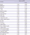
Table 2 shows correlations between baPWV and clinical parameters in total patients.
Maximal baPWV was positively correlated with mean blood pressure (r = 0.404, P < 0.001), heart rate (r = 0.285, P = 0.004), AST (r = 0.409, P < 0.001), ALT (r = 0.329, P = 0.001), γ-GTP (r = 0.273, P = 0.006), urine albumin/creatinine ratio (r = 0.321, P = 0.003). We compared baPWV according to smoking, anti-platelet treatment, alcohol intake, and statin treatment. Smoking and statin therapy showed significant positive correlation (P = 0.033, P = 0.020).
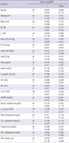
Table 3 shows correlations between baPWV and clinical variables in male and female patients respectively. Mean blood pressure, AST, ALT, γ-GTP, and triglyceride were positively correlated with maximal PWV in both groups. Heart rate and urine albumin/creatinine ratio were positively correlated with maximal baPWV only in male group (P = 0.001, P = 0.001). The c-peptide was positively correlated with maximal baPWV only in female group (P = 0.012).
Including these significant variables and well-known baPWV-related variables, we performed multivariate linear regression analyses for the association between several variables and maximal baPWV (Table 4). These analyses were performed in male, female and total group, respectively. Mean blood pressure and heart rate were significantly associated with maximal baPWV in male and total group. And in female group, mean blood pressure was the only variable associated with maximal baPWV.
DISCUSSION
In prior studies, arterial stiffness was associated with age, gender, mean blood pressure, heart rate, obesity, dyslipidemia and hyperglycemia (14, 19, 20). Those studies have been conducted in various groups such as healthy subjects and patients with impaired fasting glucose, impaired glucose tolerance, DM, chronic kidney disease and so on (17, 21-23).
To our knowledge, this study is the first to evaluate related factors to baPWV in normotensive young adults with type 2 DM. Blood pressure, age and DM are well known determinants of PWV. Therefore, we thought that it was meaningful to evaluate what factors were related to arterial stiffness in this newly selected population. We found that mean blood pressure and heart rate were significantly correlated to baPWV in male and total population of this group. Only mean blood pressure was significantly correlated to baPWV in female population.
It was noted that possible PWV-related factors such as waist circumference, body mass index, age, pulse pressure, HbA1c, ALT, γ-GTP, hsCRP, diabetes duration, albuminuria status and lipid profile did not show correlation with baPWV in this population. However, mean blood pressure and heart rate were significant predictors to baPWV.
The subjects in this study can be considered as being with short duration of diabetes and relatively healthy. Intensive complication tests are not usually indicated for these patients; and patients themselves do not realize the importance of such tests. Therefore, the finding of this study may have clinical importance in this aspect. Through easy way like measuring blood pressure and heart rate, we can predict the degree of arterial stiffness in this population like other patients group with advanced diseases.
In summary, this is the first study describing the factors influencing to baPWV in normotensive young adults with type 2 DM. The heart rate and mean blood pressure are significant influencing factors to baPWV. They can be surrogate markers of arterial stiffness in normotensive young adults with type 2 diabetes. Our study has limitation such as relatively small numbers and lack of factors of cardiovascular prognosis. For better understanding of risk factors of arterial stiffness and cardiovascular risk in this population, future large-scale studies including factors of cardiovascular prognosis are necessary.

 XML Download