

 PDF
PDF ePub
ePub Citation
 Print
Print


INTRODUCTION
Infection with human papillomavirus (HPV) is considered to be a pre-requisite for the development of cervical cancer and is associated with neoplastic precursor lesions (1). In Korean women, the cervical cancer is the fifth frequent cancer after breast cancer, stomach cancer, colon cancer and thyroid cancer. To date more than 100 HPV genotypes have been identified and at least 50 are known to infect the female anogenital tract (2). Among these thirteen mucosotropic HPVs (types 16, 18, 31, 33, 35, 39, 45, 5, 52, 56, 58, 59, and 68) have been classified as class I high risk carcinogens to human beings (3). Because the distribution and prevalence of HPV vary by geographic region and the immunity conferred by vaccines is type-specific, the need for HPV genotyping in routine screening population is increasingly recognized. For example, although HPV-16 is the most prevalent type worldwide, some genotypes such as type 52 and 58 are rare in Western countries, whereas they are relatively prevalent in Asian populations (4-6). Hence, an accurate assessment of the regional, community-based distribution of HPV genotypes is extremely important for the prevention of cervical cancer and for public hygiene management. The US FDA-approved Hybrid Capture II HPV DNA test (HC II) (Digene Corporation, Gaithersburg, MD, USA), which can detect 13 carcinogenic HPV types, is the HPV DNA detection method most commonly used. Unfortunately, this cocktail detection method does not identify specific HPV genotypes. The restriction fragmentation mass polymorphism (RFMP) assay (GeneMetrix Co., Yongin, Korea), which utilizes PCR to amplify the HPVL1 gene followed by enzyme restriction and mass measurement using MALDI-TOF, offers a method to detect 38 types of HPV. This new method has manifested sensitivity and reliability comparable to HC II (7-9).
The goal of this study was to establish and investigate an age-specific HPV prevalence, genotype distribution and extent of multiple infections in Korean women to provide the most representative epidemiologic data for the clinical prediction of the outcome of HPV infection, future prevention strategies, and the development of the efficient multivalent HPV vaccine.
MATERIALS AND METHODS
Study population
The residual samples after liquid-based cytology (LBC) tests from 60,775 patients aged 18-79 yr (median age 44-yr-olds) from September 2006 to September 2011 were used for HPV genotypes using RFMP assay. After cytology, residual cells in the LBC sample were centrifuged at 3,500 rpm for 10 min and stored as split cellular pellets of 200 µL at -70℃ prior to nucleic acid extraction.
DNA extraction
QIAamp1 DNA Blood Mini Kits (Qiagen, Valencia, CA, USA) were used to extract cervical cell DNA according to the manufacturer's protocol. Nucleic acid was stored at -70℃ prior to HPV amplification and genotyping.
HPV DNA amplification and genotyping
The presence and genotype of HPV in all samples were tested using RFMP method. The details of the RFMP assay protocol were described previously (8). Briefly, 4 µL of DNA were amplified with PGMY09/11 primers, comprising of two non degenerate pools of L1 consensus primers. Second round primer pairs comprised a sense primer RFMP specific to bases 6584 to 6603 (5'-GCMCAGGGHCAYAAGGATGAA TGG-3') and an antisense primer RFMP specific to bases 6657 to 6626 (5'-GTACTDCKDGTRGTATCHACMACGGATGTAACAAA-3'). The 5-nucleotide sequence (GGATG) embedded in the primers introduced a FokI site (a neoschizomer of BtsCI) in the amplicon. Restriction enzyme digestion of PCR products was performed by mixing the PCR reaction mixtures with 10 µL of buffer containing 50 mM potassium acetate, 20 mM Tris-acetate, 10 mM magnesium acetate, 1 mM dithiothreitol and 1 unit of FokI and BtsCI. The reaction mixtures were incubated at 37℃ for 1 hr. The resulting digest was purified by vacuum filtration through 96-well Oasis® µElution Plates (Waters, Tokyo, Japan). The desalted reaction mixtures were resuspended with matrix solution containing 15 mg/mL 3-hydroxypicolinic acid, 0.023 M ammonium citrate and 12% acetonitrile, and spotted in 3-µL volumes on a polished MTP AnchorChip™ plate (BrukerDaltonics, Bremen, Germany). Mass spectra were acquired with the aid of installed software (flexcontrol 3.0) on a Microflex linear MALDI-TOF mass spectrometer (BrukerDaltonics, Bremen, Germany).This method was able to detect 38 HPV genotypes (6, 11, 16, 18, 26, 30, 31, 33, 34, 35, 39, 40, 42, 43, 44, 45, 51, 52, 53, 54, 55, 56, 58, 59, 61, 62, 66, 68, 70, 72, 73, 74, 81, 82, 83, 84, 89, and 90).
RESULTS
HPV prevalence
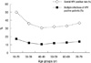
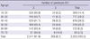
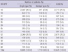
Of the 60,775 Korean women aged between 18 and 79 yr, HPV DNA was detected in 20,787 (34.2%); 10,628 (17.5%) patients were infected with high-risk HPV genotypes and 10,159 (16.7%) patients with low-risk HPV genotypes (Table 1, 2). Among HPV-positive patients of 20,878, 18,234 (87.7%) patients had single infection and 2,553 (12.3%) had multiple infection (Table 3). Age-specific HPV infection prevalence peaked at 49.9% in 18- to 29-yr-olds, and declined to 30.7% in 40- to 49-yr-olds, after which HPV prevalence steadily increased to 36.6% in 70- to 79-yr-olds (Table 1, Fig. 1). In the multiple infections of 2,553, infections with two different genotypes were 2,347 (91.9%) and with three genotypes 206 (8.1%) (Table 4). The number of genotypes was not significantly different among the different age groups (P = 0.26, Table 4). Age-specific prevalence curve of multiple infections in HPVpositive patients revealed a very similar pattern to that of the overall HPV positivity stratified by age (Fig. 1).
Age-specific HPV genotype distribution
We considered HPV genotypes 16, 18, 31, 33, 35, 39, 45, 51, 52, 56, 58, 59, and 68 as primary carcinogenic (high-risk) types and all others as non-oncogenic (low-risk) types (3).
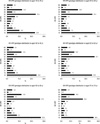
Among the high-risk HPV (HR-HPV) infections, HPV 16 (25.6%) and HPV 52 (25.2%) were the most commonly detected types, followed by HPV 58, 18, 56, 31, 51, 68, 35, 33, 45, 39, and 59 at 11.5%, 7.5%, 5.2%, 5.0%, 4.8%, 4.2%, 3.5%, 2.6%, 2.4%, 1.4%, and 1.3%, respectively (Table 5). The leading four HR-HPV genotypes stratified by age in descending order were HPV type 52, 16, 58, and 18 in 18- to 29-yr-olds, and 40- to 59-yr-olds, HPV 16, 52, 58, and 18 in 30- to 39-yr-olds, and 60- to 69-yr-olds, and HPV 16, 52, 58, 56, and 68 in 70- to 79-yr-olds (Fig. 2).
DISCUSSION
HPV is one of the most common sexually transmitted infections worldwide (10). The estimated risk of HPV is notably high over a woman's lifetime (over 80%), however, most women who acquire HPV infection do not develop more serious high-grade cervical neoplasia or invasive cancer because most infections are transient (11).
Our cross-sectional study was performed on patients referred to the gynecology office seeking for an opportunistic cancer screening with liquid based cytology. Population-based data for HPV-type distribution is a prerequisite to the development of new HPV vaccine and cancer screening tests, and to assess the effect and benefits of vaccination. The prevalence of cervical infection with human papillomavirus (HPV) in women varies geographically (12). This study represents, to our knowledge, the largest study of HPV prevalence, including HPV-DNA prevalence and a type specific distribution stratified by age in 60,775 women in Korea. Therefore, the present study provides a more closely represented HPV prevalence of the general population in Korean women for vaccine development and future prophylactic programs in Korea. The cytology results of all samples were composed of 84% of normal cytology, and 16% of abnormal cytology (data not shown).
Aside from geographical differences, variation may also be the result of detection methods employed and varied specimen type and amount. Therefore, precise and accurate HPV DNA test depends on the selection of a proper HPV genotyping method. Although the FDA-approved Hybrid Capture II (HC II) HPV DNA test is the most widely used HPV DNA detection method, it does not have the capability to identify a specific HPV genotype. Furthermore, previous studies have shown that the HC II HPV test cross-reacts with at least 15 HPV genotypes not included in its current high-risk probe cocktail set (13, 14). In this study, HPV genotypes were identified by using RFMP assay which is comparable to HC II with regard to detection of high-risk HPV infection, but is also able to identify 38 HPV genotypes in a single assay, which offers high analytical and clinical sensitivity and the advantage of reliable detection of multiple HPV infections, and it showed a good correlation with direct sequencing data (7, 9). Several studies on the prevalence of overall HPV infection in Korean women have reported rates ranging from 7.2% to 44.8%, and the prevalence of HR-HPV infection ranging from 7.2% to 34.3% (15-20). Because the prevalence of HPV infection varies by country, region within country, detection method, sample size and specimen type, the prevalence of HPV in Korean women shows in wide range according to various articles. As this study was conducted with a very large number of samples of 60,775, using validated precise and accurate detection method, the prevalence of HPV infection observed on this investigation might be the most accurate. Overall, HPV infection rate of our study was 34.2%, and high-risk HPV infection positivity was 17.5%. Those results are lower than the previous study in which overall HPV prevalence accounted for 44.8% and high-risk HPV infection rate was 34.3% (19). Age-specific HPV prevalence varied considerably across geographical regions. Across all geographical regions, observed HPV prevalence was strongly associated with age, and the curves of HPV prevalence according to age were characterized by a U-shape with a relatively higher HPV prevalence in younger and older ages in Africa, Central/South America, Europe, and North America (21). This study also revealed such U-shape curve, which peaked at women between 18- and 29-yr-olds, decline women between the age of 40- and 49-yr-olds, and then steadily increased to women between the age of 70- and 79-yr-olds (Fig. 1). The U-shaped curves of HPV by age may potentially be explained by newly acquired HPV infections and reactivation of latent HPV infections in the older women (22, 23).
Multiple infections among HPV positive cases were accounted for 12.3%, which was lower than previous study which revealed 20.9% (14). Several studies reported that the risk for developing the tissue abnormalities or lesions that typically precede cervical cancer is much higher for women infected with multiple genotypes of the human papilllomavirus. Therefore, incorporating diagnoses of multiple HPV infections into the prediction outcomes of HPV infections is considered important (24, 25). The prevalence of multiple HPV infections in HPV positive cases has shown a worldwide geographical variation ranging from about 9% to 50% in the European countries (26, 27). This variation is also due to different sample size, population, detection methods and the type of the samples.
Distribution of HPV genotypes varies across geographic areas (12, 27, 28). Several studies demonstrated that the most common type is HPV 16 followed by 18 in Europe, Central and South America, by HPV 52 and HPV-58 in Asia, by HPV-53 and HPV-52 in North America (6, 10, 27, 29). These differences in HPV type distribution in countries and regions may be related to different sexual habits and migrations of people (27, 30). Therefore, accurate information on the distribution of HPV genotypes based on the population is very important for both primary cervical cancer screening and prophylactic vaccination policy decision making.
Data on the distribution of HPV genotypes in Korean women remain controversial. Cho et al. showed that the three major HPV types were HPV 16, 18, and 33 (17). Shin et al. showed that the most common HPV types in southern Korean women were 70, 16, and 33 (20). Hwang et al. reported that three major HPV types were HPV-16, HPV-52, and HPV-58 in descending order, which is the same result of our study. The study by Hwang et al. is based on relatively large sample size of 2,470 (19).
Our study showed that the five most prevalent high-risk HPV types were Type 16, 52, 58, 18, and 56 in descending orders. Those five HPV types comprised about 76.7%, and four most prevalent types comprised 72.0% of all the 13 high risk HPV type infections. On the other hand, HPV-16 and 18, which was the most common HPV type in the western countries, comprised 34.0% of all the 13 high risk HPV type infections. The four most prevalent high-risk HPV types were not different across six age groups, except the fourth prevalent high-risk HPV type was HPV-56 and 68 in women between aged 70- and 79-yr-olds. Therefore, high risk HPV types other than HPV-16 and 18 such as genotypes 52, 58, and 56 must be targeted in the design of diagnostic tests and prophylactic vaccines for Korean women.
HPV genotype prevalence and distribution were different compared with the other regions, showing higher frequencies of HPV 52, and HPV 58. The rate of multiple genotype infections was lower than other countries as 12.3%. The most common four HR-HPV genotypes were HPV16, 52, 58, and 18 in descending order. There is a peak in HPV prevalence among women less than 30 yr of age, with a second peak among older females aged 70 to 79 yr in Korea.
In conclusion, this study provides the most representative prevalence and type-specific distribution of HPV among Korean women, and demonstrates that the epidemiology of HPV infection is different from that of other world regions.

 XML Download