

 PDF
PDF ePub
ePub Citation
 Print
Print


INTRODUCTION
Bullae and sweat gland necrosis are rare clinical and pathological entities that are associated with drug-induced coma and carbon monoxide poisoning (1, 2). Skin lesions, including bullae, purplish plaques, and erosions occur on the extremities and trunk, especially at pressure points (1, 2). The prominent histopathological features include necrosis of the eccrine secretory coils (3).
In this study, we report the case of a patient with clinical and histopathological findings characteristic of bullae and sweat gland necrosis that developed due to heavy alcohol consumption. Although this condition was first thought to be caused by Vibrio vulnificus infection or cellulitis, the subsequent clinical course and histological findings of the patient confirmed the diagnosis of bullae and sweat gland necrosis.
CASE DESCRIPTION
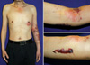
A 46-yr-old man with a history of chronic alcohol consumption for the past 20 yr presented with erythematous bullous lesions on the left arm (Fig. 1) on April 20, 2010. The patient reported that after the ingestion of clams and alcohol, he fell asleep in a state of intoxication for 12 hr with unconsciousness. After he awoke, he noticed skin lesions that first appeared as reddish patch that subsequently became hemorrhagic bullae. A dermatological examination showed multiple broad-based tense bullae and erosions on the left arm with prominent edema (Fig. 1). Two erythematous plaques were also observed on the left upper arm and chest which produced a mild heating sensation (Fig. 1). At the initial examination, the patient had mild tenderness and stiffness in the left arm, and also complained of decreased grasping power and paresthesia of the hand. He denied any recent trauma to the arm or any other part of the body.
V. vulnificus infection, cellulitis, and necrotizing fasciitis were considered in the differential diagnosis due to the patient's personal history of chronic alcoholism, ingestion of raw seafood, and the findings of hemorrhagic bullae, and paresthesia of the hand. The patient was immediately referred to the emergency room for further evaluation. His body temperature was 36℃, and other vital signs were within normal ranges. The initial laboratory findings included a leukocyte count of 6.49 × 10⁹/L and a C-reactive protein level of 1.18 mg/dL with normal hemoglobin, hematocrit, and platelet counts. A liver function test showed the following values: aspartate aminotransferase, 173 IU/L; alanine aminotransferase, 57 IU/L; alkaline phosphatase, 579 IU/L; lactic dehydrogenase, 535 IU/L; and total bilirubin, 0.88 mg/dL. Titers were negative for hepatitis A and B, and the human immunodeficiency virus.
To identify possible bacterial infections, consecutive cultures and gram stains of the blood, urine, and tissue were carried out. An MRI of the left arm revealed diffuse soft tissue enhancement on the proximal medial side and distal posterolateral areas of the left arm, suggesting cellulitis and edema. The hand and forearm radiographs did not reveal any remarkable abnormal findings.
On the basis of suspected cellulitis, intravenous ceftriaxone (2 g daily) therapy was initiated. During the remainder of the hospitalization, no new tense bullae or erythematous plaques developed. Two weeks later, the skin lesions appeared to be completely healed with only mild erythema.
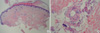
A skin biopsy from the erythematous nodule on the upper arm revealed focal epidermal necrosis with re-epithelialization and a mild perivascular lymphohistiocytic inflammatory cell infiltrate (Fig. 2A). In the dermis, there was extensive necrosis of the eccrine secretory coils and eccrine duct, and eosinophilic homogenization of the cytoplasm (Fig. 2B). In addition, bacterial cultures from the blood, urine, and tissue were all negative for pathogens. Therefore, both V. vulnificus infections and cellulitis were ruled out. Overall, the clinical course, microbiological and histopathological results were consistent with the diagnosis of bullae and sweat gland necrosis.
DISCUSSION
Bullae and sweat gland necrosis arise rare cutaneous manifestation associated with prolonged impairment of consciousness (1, 2, 4). The characteristic features of sweat gland necrosis were first documented in a patient in barbiturate-induced coma (2). Similar findings have been reported in patients who are comatose due to drug overdose, those with neurological or metabolic disorders (e.g., cerebral tumor, cerebrovascular accident, head injury, viral encephalitis, hypoglycemia, and diabetic ketoacidosis), and in immobilized non-comatose patients (2). Although barbiturates remain the most frequently reported drug associated with these entities, other causative agents include benzodiazepines (2, 3, 5), narcotics (6, 7), tricyclic antidepressants (8), and alcohol (9).
Clinically, bullae and sweat gland necrosis are characterized by bullae, violaceous plaques, erosions, and macular erythema. They are typically localized to the skin overlying bony prominences on the extremities and trunk (1). The skin lesions appear within one hour to several days after the ingestion of drugs, and resolve within 10-14 days (2, 10). There is no specific therapy for bullae and sweat gland necrosis and so they are typically treated with supportive care only (11).
The pathogenesis of the skin changes remains unclear, although several theories exist that involve: pressure (12), hypoxia (6, 12), drug toxicity (7, 13), and immune-mediated mechanisms (14). It was initially ascribed to local pressure and hypoxia because the lesions are commonly noted over a bony prominence (12). More significantly, the secretary portion of sweat glands may be particularly sensitive to such hypoxic damage (12). However, this does not explain the occurrence of similar lesions that are not associated with trauma or that are located at pressure prone sites. Others have suggested a direct toxicity related to specific drugs as important factors in the pathophysiology of this skin disorder (13). However, similar findings are also seen in nondrug-induced comas (15). To date, the cause of this skin disorder remains to be determined, and several factors such as pressure, hypoxia, and trauma are most likely to be associated with this disorder (2, 3).
In the current case, the histopathological features were characterized mainly by necrotic changes of the eccrine secretory unit (the eccrine secretory coils and eccrine duct) and bullous lesions with necrotic epithelium. The dermal blood vessels exhibited with mild degenerative changes, erythrocyte extravasation, and a slight perivascular inflammatory infiltrate of the lymphocytes (1, 2).
In the case reported here, the patient's clinical features and past history suggested several dermatoses for differential diagnoses, including V. vulnificus infection, cellulitis, and necrotizing fasciitis.
First, hemorrhagic bullae and past history of seafood intake in chronic alcoholic requires careful examinations in order to rule out V. vulnificus infection. This is because the infection can cause fulminant cellulitis, myositis, necrotizing fasciitis, and death. Vibrio cellulitis is painful, and has a rapid onset within 12 to 24 hr of exposure. In septic patients, large hemorrhagic bullae commonly arise on the extremities or trunk and usually progress to necrotic ulcers and necrotizing fasciitis (16). Nevertheless, cutaneous manifestations of V. vulnificus infections were observed in various cutaneous manifestations such as bullae, pustules, petechiae, purpura, papules, macules, cellulitis, urticaria, and erythema multiforme-like lesions (17). Therefore, the findings of bullous lesions in alcoholic patients with a recent history of exposure to sea water or the ingestion of raw seafood should alert the physician to the possibility of V. vulnificus infection. Although our patient has very similar cutaneous lesions, the clinical course and absence of systemic symptoms allowed V. vulnificus infection to be ruled out. In V. vulnificus infection, before extensive cutaneous lesions appear primary septicemia often begins with prodromal symptoms including watery diarrhea, fever, chills, nausea, vomiting, and abdominal pain (18). In addition, the sepsis has a rapid progression, and most of patients are in shock with hypotension at the time of hospital arrival (18).
Second, it was considered that the patient had cellulitis because of the clinical features and the results from the radiographic and MRI evaluations. Usually, the diagnosis of cellulitis is made by clinical features, and it often presents as erythematous patch with ill-defined, non-palpable border (16). Our patient has unique clinical features with extensive healing erosions and hemorrhagic bullae, which prevented an easy diagnosis of cellulitis. However, in some cases of cellulitis, it is possible that the overlying epidermis undergo bullae formation or necrosis (16). Therefore, a clear differentiation between bullae and sweat gland necrosis and cellulitis can be difficult until histologic confirmations. In this case, chronic alcoholic with bullae and a previous history of coma can be a clue to the diagnosis of bullae and sweat gland necrosis.
Last, the diagnosis of necrotizing fasciitis, which could be made because of the patient's history and examinations, was supported by leukocytosis, soft-tissue gas on radiographs, positive blood cultures, and deteriorating metabolic and hemodynamic status (16). However, the absence of typical radiologic and hematologic findings allowed necrotizing fasciitis to be ruled out.
In summary, bullae and sweat gland necrosis associated with alcoholism are rare conditions but should be considered in patients with bullae and a previous history of coma. It is important to maintain a high level of suspicion and recognize the characteristic histological findings in the diagnosis of this peculiar skin disorder.
 XML Download