

 PDF
PDF ePub
ePub Citation
 Print
Print


INTRODUCTION
Corticotrophin-releasing factor (CRF) plays a major role in coordinating stress responses (1). The CRF system has been implicated in the pathophysiologies of anxiety and depressive disorders, chronic pain and fatigue states, sleep disorders, acute and chronic neurodegeneration, allergic and autoimmune inflammatory disorders, and metabolic syndrome (1-3). CRF administration mimics stress responses in the gastrointestinal tract. Moreover, both peripheral and central injections of CRF receptor antagonist can inhibit stress-induced changes in intestinal function (4, 5). Thus, responses of the gastrointestinal tract to stress are presumed to be mediated by CRF (4, 6). On the other hand, colonic responses to immobilization stress have been reported to be related to mast cell degranulation (1). Mast cell tryptase activates proteinase-activated receptor-2 (PAR2), which subsequently enhances paracellular permeability (2). Increased mucosal permeability may induce motor and sensory abnormalities in the colon. Recently, we found that CRF is involved in these colonic responses to stress (7).
In the rat esophagus, acute stress can dilate mucosal intercellular spaces (6). This dilation increases mucosal permeability, which may induce motor and sensory dysfunction of the esophagus. Accordingly, stress seems to be involved in the pathophysiology of reflux-related symptoms and/or mucosal damage via the dilation of esophageal intercellular spaces.
We hypothesized that CRF played a crucial role in stress-induced dilation of intercellular spaces in esophageal mucosa. To verify this hypothesis, we examined whether blocking endogenous CRF activity, using astressin (a nonspecific CRF receptor antagonist), can prevent the stress-induced dilation of intercellular spaces in esophageal mucosa.
MATERIALS AND METHODS
Animals
Eighteen adult male Wistar rats (250-300 g; aged 14-20 weeks) were used in this study. All rats were acclimated for 7 days before experiments and allowed free access to food and water. The animals were maintained under a 12-hr light:dark cycle and isolated from environmental stressors (noise) as much as possible. The animals were housed in pairs in cages and kept in a temperature-controlled room (21 ± 1℃). All protocols were approved by the Institutional Animal Care and Use Committee of Ajou University School of Medicine (AMC-69).
Experimental protocols
The rats were handled daily for a week by the same examiner and then submitted to restraint stress or sham stress for 90 min. During all stress sessions, rats were immobilized by placing them in Plexiglass cylindrical restrainers. Rats were divided into 3 experimental groups (6 rats per group) as follow; 1) the non-stressed group (rats were injected with saline 0.1 mL intravenously and then placed freely in their home cage for 90 min), 2) the stressed group (rats were injected with saline 0.1 mL intravenously and then placed into the restraint tube for 90 min), 3) the astressin group (rats were injected with astressin, a nonspecific CRF receptor antagonist [20 µg/kg in 0.1 mL] intravenously and then placed into the restraint tube for 90 min). During all stress sessions, the total body of the animal from head to lower hind limbs was tightly placed in a Plexiglass cylindrical restrainer for immobilization. Control rats were placed in their home cages for 90 min without exposure to any restraint stress. Immediately after completing the experiments according to the protocol, all rats were sacrificed by stunning and posterior exsanguination. Blood and tissues were collected immediately after sacrifice. Astressin was purchased from the Sigma Chemical Company (St. Louis, MO, USA).
Histological evaluations
Biopsied mucosal tissues from the esophagus were fixed in formalin immediately after being removed, and embedded in paraffin wax. Serial sections were stained with hematoxylin and eosin for routine histological evaluation under light microscopy.
Measurement of intercellular space diameters
One segment (0.5 cm) of the distal esophagus was excised and fixed in 2% glutaraldehyde in phosphate buffer for transmission electron microscopy (TEM). Briefly, specimens were prepared by rinsing in buffer, post-fixing in 1% buffered osmium tetroxide at 4℃, and by dehydrating them through a graded alcohol series. They were then infiltrated with propylene oxide and embedded in an epoxy resin. Ultrathin sections on copper grids were post-stained with uranyl acetate and lead citrate. Each specimen was then examined and photographed using a Zeiss transmission electron microscope (Zeiss, Oberkochen, Germany). Three TEM photos/per rat, showing a whole cell with opposite cell membranes, were randomly taken (× 4,000 magnification) and analyzed using image analysis software in Image-Pro PLUS ver. 4.5 software (Media Cybernetics, Silver Spring, USA) (Fig. 1). Photos were stored on a disk. On each of the TEM photos, 10 transect lines were drawn across selected areas of intercellular spaces to obtain 30 transects available for measurement from each rat. The diameter of each of the transected intercellular spaces was then determined with internal scale markers. Intercellular space diameters of each rat were assessed by a pathologist unaware of the group to which the individual rat belonged.
Measurement of plasma cortisol levels
Blood samples from the central vein were collected in heparinized Eppendorf tubes immediately after sacrifice, and subsequently centrifuged (10,000 rpm, 1 min at 4℃) to obtain plasma, which was then stored at -70℃ until required for assay. Plasma cortisol levels were quantified using a radioimmunoassay kit (Rat Corticosteroid Coat-a-Count Kit, Diagnostic Products Corp., Los Angeles, CA, USA).
Statistical analysis
All data are expressed as means ± SEM. Intercellular space diameters and plasma cortisol levels were compared between the three groups by one-way ANOVA. Comparisons between two groups were performed using the Student's t-test. P values of < 0.05 were considered statistically significant. SPSS for Windows version 11 (SPSS Inc., Chicago, IL, USA) was used for all analyses.
RESULTS
Histological findings and intercellular space diameters
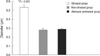
No gross inflammation or erosive lesion of esophageal tissues was observed in any rat. Under light microscopy, no histological evidence of inflammation, such as, inflammatory cell infiltration, was observed in the esophageal mucosa of any of the three study groups. The mean intercellular space diameter in the saline-pretreated stressed group was significantly greater than in the non-stressed group (0.53 ± 0.03 µm vs 0.28 ± 0.02 µm; P < 0.001). The mean intercellular space diameter in the astressin-pretreated stressed group was significantly lower than in the stressed group (0.29 ± 0.01 µm vs 0.53 ± 0.03 µm; P < 0.001). The mean intercellular space diameters in the non-stressed and astressin-pretreated stressed groups were similar (Fig. 2).
Plasma cortisol levels
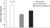
Plasma cortisol levels in the stressed group were significantly higher than in the non-stressed group (4.2 ± 0.4 vs 2.5 ± 0.4 µg/dL; P < 0.05). Plasma cortisol levels tended to be lower in the astressin-pretreated stressed group than in the saline-pretreated stressed group (3.1 ± 0.7 vs 4.2 ± 0.4 µg/dL; P = 0.08) (Fig. 3).
DISCUSSION
The present study confirms that acute stress provokes intercellular space dilation in esophageal mucosa. In addition, our data show for the first time that pretreatment with astressin, a nonspecific CRF antagonist, can prevent stress-induced alterations in esophageal intercellular spaces. Given that endogenous CRF activity is blocked by a CRF receptor antagonist, our observations suggest that CRF plays a mediating role in this stress response. Intercellular space dilation of esophageal mucosa has been reported to be involved in the pathophysiology of gastroesophageal reflux disease (GERD) (8-10). Accordingly, CRF appears to have relevance in the pathophysiologic mechanism of GERD.
As compared with other areas in the gastrointestinal tract, esophageal mucosa is less permeable to the passage of molecules (11). Dilation of intercellular spaces in esophageal mucosa may allow acid to access sensory nerve endings in esophageal wall, and cause heartburn (11, 12). A previous study has already shown that acute stress can provoke intercellular space dilation and increase mucosal permeability in the esophagus (6). These alterations can allow acid and/or pepsin to reach mucosal chemoreceptors, and thus, contribute to the genesis of reflux-related symptoms. Actually, stressful life events can induce the symptoms of GERD and increase the severity of heartburn (13, 14). Likewise, acute laboratory stress has been reported to increase sensitivity to esophageal acid exposure in patients with erosive or non-erosive reflux disease (NERD) (15). Dilated intercellular spaces in esophageal mucosa represent increased mucosal permeability to refluxed materials including acid and pepsin, which may be responsible for the activation of sensory and motor neurons. These alterations can lead to enhanced motility and sensitivity in the esophagus. Therefore, even small amount of acid refluxate may generate symptoms such as heartburn and chest pain. Intra-patient intercellular space diameters are stable over time, not overlapping with those of controls, indicating dilated intercellular spaces of esophageal mucosa are time-reproducible (10). Actually, intercellular space dilation in esophageal mucosa has been suggested to be an objective, structural marker of GERD.
Dilated intercellular spaces in esophageal mucosa of GERD patients might be the result of tissue injury due to the reflux of gastric contents. However, given that NERD patients who have not been exposed to esophageal acid at pathological levels also can show significant dilation of intercellular spaces in the esophageal mucosa, this intercellular space dilation is more likely to be attributed to mechanisms other than tissue injury by esophageal acid exposure (8). Recent studies have demonstrated that stress may provoke intercellular space dilation and a permeability defect in esophageal mucosa (6).
The implication of hypothalamic-pituitary-adrenal axis in stress-induced changes of the gastrointestinal tract has been comprehensively studied. Studies reveal that CRF is the main neuroendocrine factor mediating the effects of stress (5, 7). CRF interacts with CRF subtype 1 and/or subtype 2 receptors, located both centrally and peripherally (16). Peripheral administration of CRF mimics stress-induced colonic mucosal and epithelial abnormalities in rats (17, 18). In addition, pretreatment of rats with the nonselective CRF antagonist inhibits the effects of acute stress on gut function (18). Based on those findings, we hypothesized that CRF might play a crucial role in stress-provoked dilation of esophageal intercellular spaces. Astressin is a specific nonselective CRF1/CRF2 receptor antagonists, and is more potent and longer acting than α-helical CRF (19). CRF1 receptor antagonists have been shown to have therapeutic potential for ameliorating stress responses, including endocrine (hypothalamic-pituitary-adrenal hormone release) (20), behavioral (development of anxiety and depression) (21), and autonomic (activation of sympathetic and sacral parasympathetic outflow) responses (22). Furthermore, it has been suggested that the CRF1 signaling pathway in the brain and the gut may be implicated in the comorbidity of anxiety/depression and diarrhea-predominant irritable bowel syndrome (23). Evidence also supports the role of brain CRF1 receptors in colonic motor and sensory responses to stress (24). Although overactive CRF1 signaling pathways are considered to be related to hypersensitivity to colorectal distension, the subtype of CRF involved in hypersensitivity to esophageal distension remains to be elucidated. CRF has much higher binding affinity to CRF1 receptors than to CRF2 receptors (25). CRF1 is considered to have roles in the central and enteric nervous systems and be related to the anxiogenic actions of CRF. Thus, CRF1 antagonists appear to be potentially useful for the treatment of stress-related alterations in the gastrointestinal tract. Since we used astressin, a non-selective CRF antagonist in the present study, we could not determine which CRF receptor subtype was involved in stress-induced dilation of intercellular spaces in esophageal mucosa. Accordingly, further investigations on CRF receptor subtypes involved in the stress effect on esophageal intercellular spaces are required.
Stress models are diverse according to the nature of the stressor and the duration of the exposure to the stressor. The experimental model of restraint used in the present study involves elements of physical stress in addition to psychological stress. Restraint stress has been used in visceral hypersensitivity and intestinal permeability studies (26, 27). A partial restraint stress model, in which the animal's fore-shoulders, upper fore-limbs and thoracic trunk are restricted for 2 hr, was reported to increase plasma levels of adrenocorticotropic hormone and cortisone, indicating activation of the hypothalamic-pituitary-adrenal axis (28). We presumed that total restraint would have a greater effect, and thus, we applied total restraint stress for 90 min. The present study confirms that our model of restraint stress significantly increased plasma cortisol levels, which suggests the activation of the hypothalamic-pituitary-adrenal axis by restraint stress. In addition, we found that pretreatment with astressin tended to reduce stress-induced increases in plasma cortisol levels.
The detailed mechanism responsible for intercellular space dilation in esophageal mucosa by CRF remains to be determined. CRF released during immobilization stress has been reported to increase mast cell numbers and mucosal permeability in the colon (7, 29, 30). Likewise, CRF might increase mast cell numbers in the esophagus, and these might be responsible for intercellular space dilation in esophageal mucosa. The mechanism underlying CRF-induced dilation of esophageal intercellular spaces warrants further investigation.
In conclusion, the present study showed that acute stress in rats enlarged intercellular spaces of esophageal mucosa, and this stress-induced alteration was mediated by CRF. Our results suggest that CRF may play a role in the pathophysiology of reflux-induced symptoms or mucosal damage.

 XML Download