

 PDF
PDF ePub
ePub Citation
 Print
Print


INTRODUCTION
Ischemic mitral regurgitation (MR) has an incidence of 10% to 20% in patients with coronary artery disease and is associated with poor prognosis (1). The pathophysiology of ischemic MR is complex and is related to asymmetric alterations in annular and ventricular geometry and function (2). The most common surgical treatment is restrictive annuloplasty to increase mitral leaflet coaptation by reducing mitral annular diameter (3). However, recent data show that this procedure is associated with a 10% to 20% rate of persistent MR soon after operation and a 50% to 70% rate of recurrent MR at five years. Furthermore, the presence of persistent or recurrent MR is associated with higher incidences of heart failure and high mortality (4).
Flexible, rigid, partial, and complete rings have been used for mitral annuloplasty in order to reduce the recurrence of MR and several ventricular procedures focusing on the papillary muscles and chorda tendineae have been performed (5, 6). Currently, no method has proven successful, although some investigators reported good results. Coronary artery bypass surgery with posterior mitral annuloplasty using a vascular strip according to the location of leaflet tethering for ischemic MR have been performed since 2001 at our institution.
The aim of this study was to investigate the clinical outcomes of posterior mitral annuloplasty using a vascular strip including changes of echocardiographic parameters and to identify predictors associated with repair failure after the procedure.
MATERIALS AND METHODS
Patients
Between September 2001 and September 2010, 96 patients underwent coronary artery bypass combined with posterior mitral annuloplasty using a vascular strip for chronic ischemic MR. Chronic ischemic MR in the present study was defined as MR lasting two weeks after myocardial infarction with left ventricular segmental wall motion abnormalities, significant coronary artery disease in the territory supplying the wall motion abnormality, and structurally normal mitral leaflets and chordae. Patients with organic mitral valve disease such as prolapse, rheumatic disease, or endocarditis were excluded. All patients had coronary artery disease and at least moderate functional ischemic MR. The severity of MR had been documented by preoperative transthoracic echocardiography and confirmed by intraoperative transesophageal echocardiography under general anesthesia.
We retrospectively reviewed the data that were prospectively collected in a computerized database. Patients were stratified into two groups based on left ventricular ejection fraction (LVEF): group I, n = 50, with LVEF > 35% and group II, n = 46, with LVEF ≤ 35%.
Operative technique
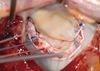
All operations were performed through standard median sternotomy. Cardiopulmonary bypass was performed with bicaval cannulation under mild hypothermia. Myocardial protection was achieved with retrograde cold blood cardioplegia. The mitral valve was exposed via a left atriotomy through the interatrial groove. Once the mitral valve was determined to be anatomically normal and the tethered part was identified, the vascular strip was prepared from Dacron artificial vascular graft according to the size of the annulus measured from trigone to trigone using Carpentier Edward ring sizer. Then, the stitches for the posterior annuloplasty were placed (usually nine overlapping 2-0 Ticron sutures without Teflon felt). Stitches were anchored at the vascular strip with consideration for the site of leaflet tethering. Because of the asymmetrical dilatation caused by the leaflet tethering (usually in the P3 segment), particular attention was paid to ensure that the middle of the vascular strip corresponded to the middle of the posterior annulus. After placement of vascular strip, we reinforced the sutures at both commissures and the middle of posterior annulus with 5-0 polypropylene in all cases to prevent from dehiscence of vascular strip. Mitral valve repair was considered successful if there was no or trivial residual MR (Fig. 1). We thought restrictive posterior annuloplasty could be effective when complete revascularization was performed concomitantly. After finishing mitral valve repair, coronary artery bypass grafting was performed in all patients. All patients underwent complete coronary revascularization. We performed left internal thoracic artery to left anterior descending artery grafting in most patients (87/96, 90.6%). The right internal thoracic artery was harvested and used as a Y composite graft to the left internal thoracic artery in 72 patients (75.0%). The in situ right gastroepiploic artery was used in 17 patients (17.7%) for revascularization for the right coronary territory. The saphenous vein was used as an aortocoronary bypass in 28 patients (29.2%) when the right internal thoracic artery was too short to reach the right coronary territory or the right gastroepiploic artery was unusable.
Echocardiographic evaluation
Left ventricular end-systolic and diastolic dimensions were determined in the parasternal long axis view at the base per American Society of Echocardiography guidelines (7). The MR severity was determined based on the ratio of maximum regurgitant jet area to left atrial area on color Doppler imaging. MR grade was graded as trivial (1+), mild (2+), moderate (3+), or severe (4+) for ratios of 0% to 10%, 10% to 20%, 20% to 40%, and greater than 40%, respectively. Recurrence of MR was defined as 2+/4+ or greater MR at postoperative time on color-flow Doppler analysis. Peak early (E) and late (A) diastolic velocities and mitral inflow deceleration time were measured using pulse-wave Doppler scanning at the tip of the mitral valve leaflets. Peak early (E') and late (A') diastolic mitral annular velocities were acquired at the septum in the apical four-chamber view. Tricuspid regurgitation (TR) was graded from 1 to 4 corresponding to mild, moderate, and severe regurgitation respectively.
Follow-up
Early mortality was defined as death within 30 days after operation or before discharge. Postoperative events after discharge were acquired from medical records, direct telephone interviews with patients or their families, or the national registry of birth and death data. Cause of death was classified as cardiac (sudden death, heart failure, or myocardial infarction) or noncardiac. Cardiovascular morbidity was defined as heart failure events, endocarditis, hemorrhage, or neurologic complications related to thromboembolism. Heart failure events included admission for heart failure symptoms, an early hospital visit and the need for more cardiac drugs such as diuretics. Follow-up was ended on September 30, 2010, and was completed in 100% of patients. The median follow-up duration was 47.9 ± 32.5 (maximum, 8.6 yr) months.
Statistical analysis
Statistical analysis was performed using SPSS ver. 17.0 (SPSS, Chicago, IL, USA). Measurements are expressed as mean ± standard deviation or as frequency and proportion. Inter-group comparisons were performed using the Student's unpaired t-test for continuous variables or the chi-squared test (Pearson's χ2 and Fisher's exact tests) for categorical variables. Repeated measures ANOVA was used to compare values at different time points. Linear regression analysis was used to detect correlations between variables of interest. Cox regression analyses were performed to identify predictors of mortality and MR recurrence. Variables assessed via univariate analysis were age, gender, diabetes, hypertension, chronic renal failure, NYHA functional class, stroke history, myocardial infarction, left main disease, emergency, atrial fibrillation, number of bypass grafts, use of saphenous vein, and echocardiographic parameters (preoperative and late follow-up). Variables with P values < 0.2 on univariate analysis were included in the multivariate analysis model. Postoperative outcomes were analyzed using the curve derived from the Kaplan-Meier method, and an inter-group comparisons were made using the log-rank test. P values < 0.05 were considered statistically significant.
RESULTS
Baseline characteristics
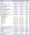
The baseline and surgical characteristics were compared between the two groups (Table 1). There were differences in baseline NYHA class, left ventricular end systolic and diastolic dimensions, and LVEF. Group II patients had a longer bypass time (P = 0.004) and the saphenous vein was more frequently used during coronary artery bypass in group II (P = 0.014).
Clinical follow-up
The early mortality rate was 2.1% (2/96). Both deaths occurred in group II, and the cause of death was low cardiac output syndrome in both cases. There were no significant differences in the incidence of perioperative morbidities such as atrial fibrillation, bleeding reoperation, neurologic complications or respiratory complication. The incidences of acute kidney injury and postoperative low cardiac output requiring prolonged inotropics longer than three days were higher in group II, the patients of which also experienced longer hospital stay. No reoperations for residual MR were performed in either group (Table 2).
There were 11 late cardiac deaths (11.5%). In group I, cardiac mortality occurred in seven patients (14%, 7/50), and the causes of death included myocardial infarction, congestive heart failure related to ischemic cardiomyopathy and sudden death. In group II, three patients died of congestive heart failure related to ischemic cardiomyopathy and one patient died suddenly (8.7%, 4/46, Table 2). The late morbidity included stroke in one patient, arrhythmia requiring hospital admission in two patients and heart failure related to moderate to severe MR in five patients. No vascular strip related complications such as detachment, hemolysis, infective endocarditis or pannus formation occurred. During follow-up, the graft patency was evaluated in 24 patients with coronary angiography or computed tomographic coronary angiography. Graft occlusion was not found in these patients, but percutaneous coronary intervention (PCI) was performed in one patient for disease progression in the native coronary artery.
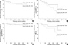
The overall cumulative survival rate at eight years was 72.9% ± 5.5% and there was no intergroup difference (77.3% ± 6.8%, group I vs 67.5% ± 8.8%, group II; P = 0.438). The proportion of patients free from cardiac-related mortality at eight years was 79.1% ± 4.9%, and there was no intergroup difference (Fig. 2A). The overall rate of freedom from mild or greater MR at eight years was 68.2% ± 7.5% and there was significant difference between groups (Fig. 2B). However, there was no intergroup difference in terms of freedom from the presence of moderate or greater MR at eight years (Fig. 2C). On the other hand, the rate of freedom from the presence of increased MR (more than one grade compared with MR at immediate postoperative period) at eight years was significantly different between groups (Fig. 2D).
Emergency operation, preoperative tricuspid regurgitation (≥ mild) and occurrence of recurrent moderate or greater MR during follow-up were predictors for cardiac-related mortality. Preoperative NYHA functional class and early postoperative residual MR ≥ mild at discharge were found to be associated with an increased risk for recurrent MR during follow-up (Table 3).
Echocardiographic follow-up
Table 4 presents the echocardiographic findings after operation. The left ventricular end systolic dimension, left ventricular end diastolic dimension and right ventricular systolic pressure were reduced during follow-up in both groups. The LVEF improved over time, and this improvement was more prominent in group II than in group I (P = 0.025). Tricuspid regurgitation was unchanged during follow-up in both groups. MR grade was reduced at discharge (0.8 ± 0.7) but increased slightly during follow-up (1.1 ± 0.8, P = 0.001). This pattern of change was not different between groups (P = 0.247).
Among the patients with preoperative moderate ischemic MR, moderate MR was detected at discharge in one patient who had mild MR on intraoperative transesophageal echocardiography after posterior annuloplasty. Forty-seven patients (47 of 63, 74.6%) had no or trivial MR during follow-up, and moderate to severe MR developed in four of 63 patients (6.3%). None of patients with preoperative severe ischemic MR had moderate MR at discharge, and 22 of 28 patients (78.6%) had no or trivial MR during follow-up. Only one patient was followed-up with recurrent moderate MR. The incidence of MR ≥ mild during follow-up was 24.2% (22/91), and there was no difference in terms of preoperative MR severity (moderate vs severe, 16/63 (25.4%) vs 6/28 (21.4%), P = 0.683, Fig. 3).
The mean transmitral pressure gradient was 3.3 ± 1.7 mmHg at discharge and 3.7 ± 2.0 mmHg during follow-up. There was no intergroup difference in the mean transmitral pressure gradient in either period (P = 0.16).
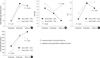
Changes in echocardiographic parameters related to left ventricular diastolic function are shown in Fig. 4. The early filling peak velocity (E) of mitral inflow increased after mitral valve repair and was maintained during follow-up in both groups. The ratio of early filling peak velocity to atrial peak velocity (E/A) of mitral inflow decreased after mitral valve repair in both groups and increased during follow-up in group II. The ratio between early diastolic mitral inflow and mitral annular velocity (E'/e) increased after mitral valve repair in both groups and slightly decreased during follow-up in group I. The mitral flow deceleration time increased after mitral valve repair in both groups and slightly decreased during follow-up in group II. However, none of the changes in echocardiographic parameters related to left ventricular diastolic function were significantly different between groups.
During follow-up, LVEF was correlated with preoperative E/A value (r = -0.255, P = 0.019), mitral inflow deceleration time (r = -0.243, P = 0.024) and left atrial volume index (r = -0.389, P = 0.009). E'/e value during follow-up was correlated with preoperative LVEF (r = -0.306, P = 0.010) and left ventricular end systolic dimension (r = 0.312, P = 0.009).
DISCUSSION
The results of our study indicate that posterior mitral annuloplasty using a vascular strip combined with coronary artery bypass is feasible and effective for treating moderate or greater ischemic MR with acceptable repair durability. There are three important points to be considered based on our findings. First, late cardiac-related mortality was not associated with the preoperative LVEF but with recurrent moderate MR during follow-up. Second, postoperative MR severity was reduced at discharge but increased during follow-up irrespective of preoperative MR severity. Third, the most important predictor of recurrent MR was early postoperative MR.
The detrimental effects of ischemic MR leading to heart failure and high mortality are well documented (1). In the past, coronary revascularization was believed to correct ischemic MR by improving left ventricular function (8). However, observational studies have demonstrated that revascularization alone does not resolve MR (9). Although some studies have reported that coronary artery bypass surgery combined with mitral valve repair shows better outcomes in terms of MR recurrence than coronary artery bypass surgery alone, controversy remains regarding operative risk and survival benefit. Grossi et al. (10) reported an early mortality rate of 13.4% in ischemic MR patients with a five-year survival of 71% after mitral valve repair. Gillinov et al. (11) found an early mortality of 13%, and Kang et al. (12) reported an operative mortality of 12% in Korean patients undergoing mitral valve repair. They also found that mitral valve repair was superior in patients with severe MR and did not increase operative or late mortality rate compared to coronary artery bypass surgery alone. However, in patients with moderate MR, the operative mortality rate of mitral valve repair was higher than that of coronary artery bypass surgery alone, whereas the MR improvement rates were similar for the two procedures. Yu et al. (13) reported a similar result. Recently, the early results of mitral valve repair in patients with ischemic MR have been improved over time as the mechanisms of ischemic MR have become better understood. Szalay et al. (14) reported an early mortality rate of 6.6% with an 85% survival rate at three years in 91 patients with ischemic cardiomyopathy and moderate to severe MR who underwent coronary artery bypass surgery and mitral valve repair. In the present study, early mortality occurred in two patients (2.1%), and this is comparable to the operative mortality of 2.4% for mitral valve repair reported by Calafiore et al. (15).
Poor outcomes are related to left ventricular damage as a consequence of remodeling in ischemic MR, and this damage can be successfully remedied with a relatively simple procedure performed on the mitral annulus, that is, undersized annuloplasty which was popularized by Bolling et al. (3). However, some surgical series showed that recurrent MR occurs in one third of patients during follow-up (4, 16). Recently, techniques for reducing the incidence of late recurrent MR have been attempted, including annuloplasty using different rings such as asymmetric, rigid, flexible, complete, or partial rings and procedures at the level of the left ventricle, papillary muscles or tethered leaflets (5, 6). Randomized data on the results of these repair techniques are lacking, and results of observational studies are not consistent. In our series, a vascular strip was used for restrictive posterior annuloplasty due to its many advantages including ease to handling, high cost effectiveness, semi-rigid ring effect and wide availability. The rate of freedom from moderate or greater MR at eight years was 85.1% ± 8.3% in our cohort, comparable to that reported by Wong et al. (17).
A reduction in left ventricular dimension is associated with favorable clinical outcome. Bax et al. (18) performed a serial echocardiographic assessment of left ventricular dimensions after mitral valve repair and found that left ventricular reverse remodeling is a gradual and time-dependent process. According to that report, regression of the left ventricular end systolic dimension was observed in 73% of patients and the left ventricular end diastolic dimension was reduced in 55% (less frequent). In the present study, we found that the left ventricular dimensions were diminished irrespective of LVEF, but the left ventricular dimensions during follow-up were greater in patients with LVEF above 35% than those with LVEF ≤ 35%. Patients with LVEF less than 35% in our series were characterized by greater left ventricular dimensions at baseline. Braun et al. (19) found that a preoperative left ventricular end diastolic dimension of 65 mm or less or a left ventricular end systolic dimension of 51 mm or less was predictive of reverse remodeling. These findings may suggest that severe left ventricular dilatation may be an irreversible process, and surgery should be performed before severe dilatation has occurred. We did not observe any changes in the left atrial dimension before and after mitral valve repair.
Although there is active debate as to whether mitral valve repair will affect survival, there are few data about the importance of residual or recurrent ischemic MR. Hausmann et al. (20) reported reduced survival in patients undergoing mitral valve repair with revascularization in whom MR grade I or higher remained and this result is consistent with that of our analysis. According to our study, one of the independent predictors of mortality was recurrent moderate or greater MR after surgical correction.
Previous efforts to identify predictors of recurrent MR after surgical correction have been demonstrated. The observational study by Penicka et al. (21) revealed that reliable improvement in moderate ischemic MR after isolated coronary artery bypass surgery was related to the presence of viable myocardium and the absence of dyssynchrony between papillary muscles. Braun et al. (19) found that the preoperative left ventricular dimensions were related to MR recurrence and reverse remodeling, and Kongsaerepong et al. (22) determined that higher MR severity, larger mitral annular diameter, and greater tethering area are independent predictors of mitral valve repair failure. In the present study, higher preoperative NYHA functional class and residual MR (≥ mild) were independent predictors of recurrent MR during follow-up. However, preoperative left ventricular function and dimensions were not significant on multivariate analysis. Based on these results, we can infer that complete elimination of MR during operation is important in preventing MR recurrence despite poor left ventricular function and dilated left ventricular dimensions.
In terms of operative technique of mitral annuloplasty using a vascular strip, no vascular strip related complications occurred and no reoperation for MR was performed. Five patients had moderate or severe MR during follow-up and the cause was tethering of leaflet due to the left ventricular dilatation in all patients. It seems that mitral annuloplasty using a vascular strip is not associated with MR aggravation in our series. However, further studies are required to reveal the correlation between the operative technique using vascular strips and MR aggravation.
Some authors have performed an aggressively undersized annuloplasty in order to reduce the incidence of recurrent MR and have achieved excellent mitral valve competence (3). However, concerns have also been raised with regard to the potential for an aggressive annuloplasty to lead to increased transmitral pressure gradients, which could have a deleterious effect on clinical outcomes (23), and this issue remains controversial (24). In our series, the mean transmitral pressure gradient was 3.3 ± 1.7 mmHg at discharge and remained stable during follow-up (3.7 ± 1.9 mmHg, P = 0.17).
The E'/e ratio is a powerful predictor of survival in patients with ischemic heart disease (25). Bruch et al. (26) validated the use of E'/e for estimating filling pressures in patients with ischemic MR. They found that an E'/e ratio > 15 had 80% sensitivity and 100% specificity for predicting a left ventricular end-diastolic pressure ≥ 15 mmHg. In the present study, the E'/e ratio was measured in all 76 survivors with a follow-up interval of 2.5 ± 2.2 (maximum, 8.2 yr) years. The mean preoperative E'/e was 29.9 ± 14.3. While the preoperative left atrial volume index, left atrial dimension and left ventricular end diastolic dimension were not correlated with E'/e during follow-up, while preoperative LVEF and left ventricular end systolic dimension were. During follow-up, LVEF was correlated with preoperative E/A, mitral inflow deceleration time and left atrial volume index. Based on these results, we suggest that early mitral valve repair should be considered in patients with left ventricular dysfunction and dilation of the left ventricular end systolic dimension in order to relieve further left ventricular diastolic dysfunction in moderate ischemic MR.
Several limitations of the present study should be mentioned. First, this study is inherently limited by its retrospective nature. However, all patients in our series underwent the same surgical procedures: complete revascularization and mitral annuloplasty using vascular strips of the same length. Moreover, the echocardiographic follow-up protocol was homogeneous in most patients as it was performed at the same institution albeit at different intervals. We believe that data collection errors were minimized for these reasons. Second, more detailed echocardiographic data such as coaptation height, tenting area, and tethering height were not available because these were not routinely measured in the past. Inclusion of a myocardial viability assessment could likely have improved the present study. However, myocardial viability was only tested in recent patients. The third limitation is in the statistical analysis. The echocardiographic parameters were not measured at the same point in time for all patients during follow-up. Therefore, direct comparisons of the parameters at the three points in time (preoperatively, at discharge, and at follow-up) may not be appropriate. However, our focus was primarily on changes and correlations between parameters during follow-up.
In conclusion, restrictive posterior mitral annuloplasty using a vascular strip combined with coronary artery bypass is feasible and effective for treating moderate or greater ischemic MR with acceptable repair durability. It is important to avoid early postoperative residual MR. Furthermore, early mitral valve repair may be helpful for patients with left ventricular dysfunction and dilation of the left ventricular end systolic dimension.


 XML Download