

 PDF
PDF ePub
ePub Citation
 Print
Print


INTRODUCTION
The gastrocnemius and soleus muscles are important for leg functions and they form a muscular mass in the calf. Both of them show numerous variations in the origin and/or insertion. Each of the gastrocnemius tertius (GCT) (1, 2) and the accessory soleus (AS) muscles (3-6) has been reported by many authors, however, there is no report on their co-existence. Present report describes this co-existence. In this study we discussed existence of the gastrocnemius tertius and accessory soleus muscles from phylogenetical, anatomical and clinical points of view.
CASE DESCRIPTION
The bilateral gastrocnemius tertius and the accessory soleus muscles were encountered during dissection studies of a 30-yr-old Turkish male cadaver in 2002-2003 education period. Both sides of the calves were normal in every aspect except for these variant muscles. Innervation of the gastrocnemius tertius and the accessory soleus muscles were from the tibial nerve.
Left side observations
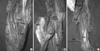
In this side, a two-bellied gastrocnemius tertius and an accessory soleus muscle were observed. The superficial belly of the gastrocnemius tertius muscle had its origin from an area just above the tendon of the plantaris muscle, and the deep belly had its origin from the tendon of the plantaris muscle itself (Fig. 1A, B). The superficial belly of the GCT inserted onto the outer surface of the lateral head of the gastrocnemius muscle after crossing the surface, the deep one inserted onto the inner surface of the medial head of the gastrocnemius muscle. The lateral and medial heads of the gastrocnemius muscle were normal in every aspect. The accessory soleus muscle originated from the posteromedial aspect of the tibia and soleal line of the tibia and inserted to the medial surface of the calcaneus independently of Achilles tendon (Fig. 1C).
Right side observations
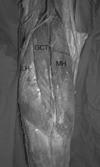
In this side, a gastrocnemius tertius muscle was observed between the lateral and medial heads of the gastrocnemius muscle (Fig. 2). It had its origin from the lateral condyle of the femur, and inserted to the medial head of the gastrocnemius muscle.
DISCUSSION
GCT is the most common variation of the gastrocnemius muscle. It was first described by Kelch (1, 5). The existence frequency of the third head was reported as 1.7%-5.5% (2, 7).
From phylogenetic point of view, the gastrocnemius muscle has been considered a muscle of the fibular side of the leg. This may also be valid for the lateral head of the soleus muscle (5). The gastrocnemius muscle comes from the calcaneum blastomere and follows an ascending migration towards the inferior femoral epiphysis. The medial head inserts higher than the lateral one, and comes into contact with the popliteal artery (8). The definition of the third head is a congenital growth of excess muscle.
The third head may arise from the linea aspera, the long head of the biceps femoris muscle, the lateral epicondyle, the knee joint capsule, the midfibula, or the crural fascia (5). It commonly joins to the medial head than to the lateral one (9). It may split and arise from more than one location or divide near its termination to join both heads of the gastrocnemius (5). In the present case, the gastrocnemius tertius consisted of two bellies in the left side and of one belly in the right side.
The absence of the lateral head of the gastrocnemius muscle and/or its reduction to a fibrous cord has been reported (5). In some cases, the two heads of the gastrocnemius muscle may be unjoined until their insertion onto the calcaneus and with separation from soleus.
The third head may be a potential problem for structures in the popliteal fossa as illustrated by Frey (10), as well as Tochihara and Onozawa (7). The third head may or may not cross the popliteal neurovascular structures. The third head joining the medial head of the gastrocnemius muscle is most commonly cited as causing clinical problems (entrapment syndromes). In some instances, the lateral head, the plantaris muscle, the accessory semimembranosus and a fibrous band may also be involved (9). The anatomical anomalies responsible for popliteal entrapment can be explained by a muscular and/or arterial embryologic development. Abnormal muscular migration of the medial head of the gastrocnemius muscle characterized by early and more or less lateral attachment to the distal femoral epiphysis leads to a more medial position for the popliteal artery in relation to the muscle (8). In most instances, the symptoms are brought by arterial occlusive changes (9) and cases which showed venous symptoms have been rarely reported (11). The characteristic signs and symptoms are: history of leg swelling, aching pain, tenderness of the popliteal fossa, which seem to be a result of chronic nerve compression. Another sign is the diminution of the pulse of the distal arteries in the passive dorsoflexion of the ankle (2). Simple Doppler examination is valuable as an initial diagnostic evaluation (12). On dorsoflexion position of the ankle, sound of the posterior tibial or dorsalis pedis arteries will be disappeared. On the other hand, venous flow evaluation rarely gives accurate confirmation (2). Surgical resection of the third head relieves the symptoms.
Although, the accessory soleus muscle was first described in 1843 as supernumerary soleus it was well documented in anatomic (5), orthopedic (6, 13) and radiological literature (3, 4).
The accessory soleus muscle may arise from the posteromedial aspect of the tibia, the anterior aspect of the Achilles tendon (6), the proximal one third of the fibula, the soleal line of the tibia, the aponeurosis of the flexor digitorum longus muscle or both the soleal line of the tibia and the anterior aspect of the soleus muscle (5). The insertion of accessory soleus is also variable. Lorentzon and Wirell (14) have described four anatomic variations of its insertion. These include: 1-insertion along the tendon of Achilles, 2-fleshy insertion to the upper surface of the calcaneus, 3-tendinous insertion to the upper surface of the calcaneus, 4-fleshy insertion to the medial surface of the calcaneus. A fifth type, tendinous insertion to the medial surface of the calcaneus, has been added by Yu and Resnick (15). In addition, it has been reported a bifid tendon inserted on the medial and lateral aspect of the calcaneus (16). In the present case, the insertion of the accessory soleus muscle was on the medial surface of the calcaneus as described by Yu and Resnick (15).
Accessory soleus muscle is a fairly uncommon congenital muscle anomaly. In the greater majority of reported cases, it exhibits a unilateral involvement (3, 6), but bilateral involvement in some reports (17). The incidence of this accessory muscle ranges from 0.7%-5.5%. It is twice as common in males as in females (6).
The accessory soleus muscle has been clinically reported in patients as young as 3 months and as old as 66 yr (at the time of diagnosis) (6), with an average age of 21 yr (14). Additionally, Peterson et al. (18) reported a 91-yr-old cadaver with accessory soleus muscle. The present case was a 30-yr-old male cadaver.
The accessory soleus muscle was considered as a variant of the plantaris muscle (13). On contrary, its fibers may originate from the deep aspect of the soleus muscle and not from the flexor muscles or plantaris muscle (5). At present, it has been postulated that the single anlage of the soleus muscle undergoes early splitting to produce an accessory soleus muscle, which may have its own blood and nerve supplies, or in common with those of the triceps surae muscle (19). Anatomically, an accessory soleus muscle is usually enclosed by its own fascia and has a tenuous blood supply from the posterior tibial artery, which might be responsible for ischemic pain following strenuous exercises (20).
The accessory soleus muscle has characteristic clinical features and radiological appearance. Lack of familiarity with these may lead to mistaken diagnosis of soft tissue tumor (4). Although gastrocnemius tertius and accessory soleus muscle have been well documented, there is no report about their co-existence. Thus this co-existence should be kept in mind during surgical procedures in the leg and ankle.
 XML Download