

 PDF
PDF ePub
ePub Citation
 Print
Print


INTRODUCTION
Congenital leukemia, defined as a leukemia that occurs within 4 to 6 weeks of birth, is rare. Excluding Down syndrome-related transient neonatal myeloproliferation, its incidence is less than 1% of all childhood leukemia (1, 2). The diagnostic criteria include the presence of immature leukemic blasts in the blood and in extrahematopoietic tissues and the absence of congenital infections (syphilis, toxoplasmosis, herpes simplex, cytomegalovirus, rubella, and bacterial infections), hypoxia, and hemolytic disease, which may produce a similar clinical and hematological picture. In addition, there may be absence of chromosomal disorders that may be associated with unstable hematopoiesis, such as trisomy 21 (3).
Although its biology and natural history are still under investigation, it is clear that the leukemic process originated in utero even in infants diagnosed within the first few months of life (2). In infants with congenital leukemia under the age of 1 month, the 6-month survival rate is only one third despite aggressive chemotherapy (4). It has a higher mortality rate than any other congenital cancer, but recently some reports showed spontaneous remissions (3, 5). These findings implicate therapeutic dilemmas on deciding which patients to treat or to wait.
Here we present a case of congenital myeloid leukemia (M5) initially characterized by the presence of leukemia cutis and hyperleukocytosis with chromosomal abnormality (double translocation of t[8;16][p11;p13.2] and t[17;19][?;q13.3]). With the fear of toxic effect of chemotherapeutic agents, parents did not agree to chemotherapy at first. Three weeks later we started chemotherapy but the prognosis was poor. We report our experience and review previous cases in view of treatment dilemmas.
CASE REPORT
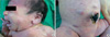
A term male infant weighing 3,360 gram was born to a 32-yr-old woman after uncomplicated pregnancy. There was no history of exposure to radiation, any known teratogens, smoking, drinking, or drug use from his mother. A cesarean section was done for previous section. Gross examination of the infant noted at birth to have an unusual purpuric rash on his whole body looking like "blueberry-muffin" rash (Fig. 1). Physical examination showed no hepatosplenomegaly and there were no dysmorphic features. Skin examination revealed widespread, scattered, yellowish-colored firm nodules with multiple purpuric macules and papules predominantly on the face and proximal extremities with a few present on the trunk. A punch-biopsy of a nodule showed proliferation of atypical hematologic cells in the dermis. Blood count at birth revealed white blood cell count of 173×109/L with 58.8% neutrophils, 5.3% lymphocytes, 26.1% monocytes, and 0.5% eosinophils; hemoglobin of 13.9 g/dL; platelets of 133×109/L. TORCH titers (against toxoplasmosis, cytomegalovirus, herpes simplex virus, rubella, and syphilis) were all negative and blood cultures were also negative. Peripheral blood morphology showed marked leukocytosis with 32% of immature cells and monocytosis. Immunophenotyping showed that blasts were positive for CD14, CD33 and HLA-DR(+). Flow cytometry on the bone marrow showed the blasts to be acute myeloid leukemia (M5) in the French-American-British classification based on the morphologic features and special stains. Bone marrow aspiration and biopsy also showed 90% of leukemic blasts. The results of special stain were peroxidase (+), PAS (-), ANAE with NaF inhibition (+). Cytogenetic analysis revealed 46, XY karyotype with multiple chromosomal defect, t(8;16)(p11;p13.2), t(17;19)(?;q13.3). Serial white blood cell count showed a progressive rise up to 256×109/L with thrombocytopenia of 88×109/L. Leukemia cutis also progressed and it became more pronounced on the face and somewhat hardened. Parents only wanted conservative treatment at first. Three weeks after birth, parents agreed to chemotherapy. Initial chemotherapy was started with daunorubicin, etoposide, and cytarabine, but, he died at 27 days of age.
DISCUSSION
Leukemia is the most common malignancy presented during the childhood, however, congenital leukemia is a very rare disease, representing less than 1% of all childhood leukemia. It should be differentiated from transient leukemoid reaction and other small round cell tumors (3).
Several risk factors are known to be associated with the development of congenital leukemia. Maternal alcohol consumption, tobacco smoking, maternal exposures to radiation, high birth weight, high levels of insulin-like growth factors, maternal consumption of topoisomerase II inhibitors, such as fruits and vegetables, coffee, tea, cocoa, wine, and soybeans are those factors (2, 6). However, as in our case, some had been reported to have no known risk factors. Frequently, reported cases of congenital leukemia with constitutional chromosomal abnormalities have led to suspicion whether congenital leukemia is a result of chromosomal fragility (2).
Congenital leukemia may reflect the early onset of intrauterine leukemia, that is, leukemic process originated in utero even in infants diagnosed within the first few months of life. Since embryonic hematopoiesis begins in undifferentiated mesenchyme starting the third week after fertilization, leukemia cutis may be a primary event and the first manifestation of congenital leukemia (6).
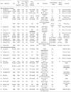
Prognosis of congenital leukemia is dismal and has a progressive downhill course, even with chemotherapy. However, there are some reported cases of spontaneous remission (Table 1) (3, 7-19). The reason that spontaneous remissions may occur in newborn infants is unclear (1). The period of spontaneous remission varied widely from months to years, and several of the children with relapses remained in prolonged remission after chemotherapy (3, 15). There appear to be no common clinical features between those who progressed and those that had spontaneous remissions. Neither bone marrow involvement at diagnosis nor hyperleukocytosis appears to increase the risk of relapse (3, 4, 15-17, 19). Chromosome abnormalities affecting chromosome 11 band q23 are involved in the majority of infant leukemia cases. The major translocations involving the 11q23 locus are t(4;11) and t(11;19) (13, 16). Translocation involving 11q23 band or reciprocal translocations involving chromosomes 8 and 16 are known to be mapped with oncogene(s) and are known to be associated with a poor prognosis (6, 10, 30). Hence, even cytogenetic abnormality of 11q23 rearrangement of skin without involvement of bone morrow was also treated aggressively (6). Moreover, AML with the t(8;16) is associated with a young age at diagnosis, myelomonocytic (FAB M4) or monocytic (FAB M5) morphology, erythrophagocytosis, disseminated intravascular coagulation and a poor outcome (9).
The gene at 11q23, known as MLL (ALL-1, HRX, HTRX, or Hu-ets-1), is required for the production of normal numbers of hematopoietic precursors and for their proper differentiation (2, 13). Chromosomal translocations can theoretically occur between any gene loci. 11q23-MLL gene rearrangements after DNA damage occur most frequently with AML. Translocations involving MLL may fuse with other genes and hence MLL usually retains at least two DNA binding domains (2, 13). Since there are a few reports of double translocations as our case, exact meaning of double gene translocation is yet to be discussed.
Because of the rarity of this disease, there is no standard protocol of chemotherapy. Treating congenital leukemia means exposure of the toxic chemotherapeutic agents to the neonate. Although there had been reported cases of prolonged periods of remission, rapidly downhill course were also noted in some cases and some of them died after relapse during the course of chemotherapy (16-18). While some cases with t(8; 16) remitted spontaneously without treatment (3, 9, 10), paradoxically, those who had chromosomal abnormalities died after initiation of chemotherapy as our case (4, 6, 20, 23, 26-29). It seems like that karyotypic findings in blasts in the neonatal period may not be predictive of whether or not a spontaneous remission will occur (2).
For our case, hyperleukocytosis, progressive leukemia cutis, bone marrow involvement, double translocation involving (8;16) made us to decide to treat at first. However, with the fear of unknown toxic effect of chemotherapeutic agents, parents refused to treat. Three weeks later after parents' agreement, we treated with daunorubicin, etoposide, and cytarabine but he died of pulmonary hemorrhage three days after induction chemotherapy. He may died of leukemia itself but, we are not sure whether he died due to chemotherapy or he could have survived with only conservative management as other spontaneous remitted cases. That is because it is the first case of congenital AML with double translocation of (8;16) and (17;19) to be reported in the English literature to the best of our knowledge.
Although congenital leukemia remains a rare disorder, an international collection of data or register system is indispensable for establishing an optimal treatment protocols.
 XML Download