

 PDF
PDF ePub
ePub Citation
 Print
Print


INTRODUCTION
Left transvenous pectoral implantation with an active Can has become the standard for implantable cardioverter defibrillator (ICD) device placement to secure a lower defibrillation threshold (DFT). However, some patients require implantation in the right pectoral region for a variety of reasons such as left pectoral infection, left subclavian access difficulty, left pectoral hardware, or left mastectomy (1). Right-sided pectoral implantation is feasible but may be associated with an unacceptably high DFT. In this report, we describe a case of ICD implantation in the left pectoral side, by tunneling after inserting a lead to the right subclavian vein, because of left subclavian vein obstruction.
CASE REPORT
A 56-yr-old man collapsed without chest pain after playing badminton on January 29, 2007. His friend witnessed the collapse and promptly performed cardiopulmonary resuscitation. The Emergency Rescue Service was called, and on arrival, after determining the presence of ventricular fibrillation, applied an automatic external defibrillator.
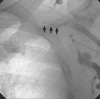
Defibrillation shock was delivered twice and sinus rhythm recovered. After admission, his mental state was markedly disoriented with retrograde amnesia. His speech was appropriate but unable to perform 7 serial subtractions. Physical examination was unremarkable. A profile of routine chemistry did not reveal any specific abnormalities except for slightly increased cardiac enzymes (CK: 1289 IU/L, CK-MB: 13.6 ng/mL) which might be due to repeated cardioversions. The electrocardiogram revealed normal sinus rhythm with left ventricular hypertrophy by voltage criteria and small Q waves in inferior leads. There was no ventricular preexcitation or QT prolongation. He had been on medication (Losartan, Thiazide, Glimepiride, Metformin) for hypertension and diabetes over the last 5 yr. According to detailed history, the patient have had exertion angina in the last 2 yr but did not seek for medical advice. The prolonged telemetry monitoring revealed an episode of non-sustained monomorphic ventricular tachycardia (8 beats, right bundle branch block morphology, cycle length 340 ms). Echocardiography and coronary angiogram were performed to evaluate the structural heart disease. An echocardiography revealed inferior wall akinesia and an ejection fraction of 42%. A coronary angiography demonstrated a chronic total occlusion of the proximal right coronary artery and patent left coronary artery without stenosis. Percutaneous coronary intervention with stent was performed in the right coronary artery without complications. During an electrophysiologic study, programmed electrical stimulation with single or double ventricular extrastimuli reproducibly induced polymorphic ventricular tachycardia with hemodynamic compromise. In view of the inducible ventricular tachycardia observed during electrophysiologic study and aborted sudden cardiac death without an acute coronary event, we decided to implant a single chamber ICD for the secondary prevention of sudden cardiac death. However, while the ICD was being implanted, a left subclavian venogram failed to visualize the left subclavian vein, and the retrograde femoral catheter could not be advanced to the vein (Fig. 1), which was attributed to likely prolonged indwelling of the left subclavian sheath for venous access. Accordingly, the right subclavian vein was punctured, and a defibrillating ventricular dual coil lead for an ICD (Vitality VR 1870, Guidant, St. Paul, MN, USA) was inserted. Lead measurements revealed ventricular sensing at 9.0 mV, a pacing threshold of 0.8 V/0.5 ms, and an impedance of 420 Ohms. Subsequently, a defibrillation test using a biphasic waveform was conducted in the dual coil system configuration (conventional configuration; RV-→SVC++CAN+).
Initially, an active Can was positioned at the right pectoral site. A DFT test of the active Can that revealed high energy (31J) to terminate the induced ventricular fibrillation. Polarity reversal, different shock configuration (RV-→CAN+) and lead repositioning were not helpful at reducing DFT (31J).
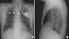
Therefore, after fixation of the lead sleeve at left pectoralis fascia, the ICD lead was diverted from the right side area to the ICD in the left pectoral area via tunnel made by tunneling tool (Medtronic, Minneapolis, MN) over the sternum. Adaptor of lead extension was not necessary because lead length was long enough. A repeat defibrillation test revealed a lower DFT of 11J, and the active Can was successfully implanted in a pocket on the left pectoral side without complication (Fig. 2). During 2 yr follow up after ICD implantation, patients had a no episode of tachyarrhythmia and recent lead measurements revealed ventricular sensing at 10.2 mV, a pacing threshold of 0.8 V/0.5 ms, and an impedance of 439 Ohms.
DISCUSSION
As the ICD pulse generator is used as one of the electrode, the active Can is routinely being placed in the left side pectoral position and the intracardiac ring electrode in RV, which theoretically includes more ventricular myocardium. Accordingly, the left subclavian route has been used for ICD lead placement. However, the subclavian vein route may sometimes be difficult to gain access to heart. In cases of critical inflammation at the left pectoral side, left subclavian vein obstruction or preexisting device at the left pectoral side, right sided route is an alternative. Gold et al. (2) reported DFT were higher on the right side and a higher mortality in patients with right sided ICDs and Ovadia et al. (3) showed that the ipsilateral supraclavicular approach is feasible and safe in cases of subclavian vein obstruction, but this approach can only be adopted after confirming the length of the obstructive segment by venography. Therefore, an alternative strategy is required when the subclavian vein is totally occluded.
Previously we reported a case (4) in which a preexisting left side pulse generator was repositioned by subcutaneous tunneling over the sternum. By using same subcutaneous tunneling technique, a permanent defibrillation lead was diverted to left pectoral side to achieve the lower DFT in this patient.
The approach used in this case might be considered in patients who had difficult left subclavicular venous access and it may be prudent to save the left subclavian vein for ICD implantation in patients with fatal tachyarrhythmia.
 XML Download