

 PDF
PDF ePub
ePub Citation
 Print
Print


INTRODUCTION
Survival rate of the patients with hepatocellular earcinoma (HCC) was increased due to the advancement of diagnosis and treatment, however, the total tendency has yet to be improved. Most cases were diagnosed at late stages. So far, the rate of surgical resection for HCC is around 20%; for most patients surgical treatment was not indicated (1). A previous study reported that the five-year survival rate after radical resection was less than 50%, and the rate of metastasis was 61.5% (2).
Tetraspanins is a large family of ubiquitously expressed membrane proteins. Several tetraspanins molecules such as CD9, CD82, CD63 and CD151 have been identified and implicated in the regulation of cell development, differentiation, proliferation, motility and tumor cell invasion (3-7). Tspan-1 (or NET-1, Gene ID: 10103) is a new member of the tetraspanins group. Sequence analysis of Tspan-1 revealed a structure typical for tetraspanins, with the presence of four transmembrane domains delimiting two extracellular regions as well as conserved amino acid residues (8). Tspan-1 was found to be overexpressed in some tumors (9-14). Studies reported Tspan-1 expression may be associated with tumor cells proliferation (9, 15). All of these reports suggest that Tspan-1 may play a critical role in the progression of tumor growth and metastasis in numerous human tumors, including HCC.
P27 is a well known negative regulator of cell cycle progression. Jab1 directly binds to p27 and induces nuclear export and subsequent degradation (16). Some reports have shown that overexpression of Jab1 and low expression of p27 is associated with advanced tumor stage and poor prognosis in several human cancers (16-19), including HCC (20).
In the present study, we examined the expression of Tspan-1 combined with Jab1, and p27 in specimens of 76 patients with HCC. The relationship between the expression of Tspan-1, Jab1 and p27 with the clinicopathological factors and the prognosis of HCC patients were explored.
MATERIALS AND METHODS
Clinical data of the patients
Seventy-six specimens in this study were obtained from the patients with HCC in our hospital undergoing surgical operations of the liver during 1998-2004. The number patients was 76 cases (male 60, female 16) and mean age was 58 yr old (from 35 to 79). Histologically 10 cases were well differentiated, 42 cases were intermediate differentiated, and 24 cases were poorly differentiated. Of them 23 cases had portal vein tumor thrombus and distant dissection. Normal control tissues of the liver were taken from ten patients operated for hemangioma. The follow-up period of the patients exceeded 60 months. Informed consent was obtained before any specimens were taken. The study protocol was approved by the Ethics Committee of the affiliated hospital of Nantong University (NTU090024).
Antibodies used in immunohistochemisty
The antibodies used for immunohistochemistry in this study were: Tspan-1 rabbit anti-human polyclonal antibody (1:100; devised by the author and prepared under the cooperation of the American San Francisco Gene Biological Company, San Francisco, CA, USA), mouse monoclonal antibody against human Jab1 (611618, 1:50;BD Biosciences PharMingen, San Diego, CA, USA), rabbit anti-human p27 polyclonal antibody (C-19, 1:50; Santa Cruz Biotechnology, Inc., Santa Cruz, CA, USA), HRP-goat anti-rabbit IgG (1:200; Sigma-Aldrich, St. Louis, MO, USA), HRP-goat anti-mouse IgG (1:200; Sigma-Aldrich, St. Louis, MO, USA).
Immunohistochemical examination of expression of Tspan-1, Jab1 and p27
Two-step immunohistochemical method was performed on formalin-fixed, paraffin-embedded 5-µm sections from all patients to detect the expression of Tspan-1, Jab1 and p27 in HCC. Five consecutive slides were prepared from each tissue. The slides were deparaffinized by dimethyl benzene, dehydrated by gradient ethanol. For antigen retrieval, they were heated at 95℃ for 10 min in sodium citrate buffer (10 mM sodium-citrate monohydrate, pH 6.0). Slides were allowed to cool for 20 min at room temperature, incubated in 0.3% H2O2 at room temperature for 15 min to inhibit endogenous peroxidase, and then incubated with primary antibodies overnight at 4℃. Two-step reagent kit (HRP- anti-mouse/rabbit IgG) was then applied to detect the immunoreactivity. Slides were stained by DAB, and counterstained by hematoxylin. Rabbit IgG was used to replace the primary antibody as control staining. Gastric cancer tissue was used as positive control.
For microscopy, five random high-magnification fields (×400) were selected from each slide to count 1,500 tumor cells. Percentage of positive cells was calculated and recorded as Label Index (LI=% of positive cells). The mean value of LI of Tspan-1, Jab1 and p27 (0.70, 0.69 and 0.26, respectively) in HCC tissues was used to define the over and low expression according to a previous study by other author (21).
Statistical analysis
The data of the expression of the proteins are presented as means±SD. Spearman test was applied to detect the correlation of among Tspan-1, Jab1 and p27 expression. Fisher's exact test was used to compare the expression of all proteins as groups (positive vs. negative) with various clinicopathological parameters. Survival analysis was undertaken using Kaplan-Meier method and group differences in survival time were investigated by log-rank test. The multiple factor survival analysis was evaluated using Cox's proportional hazards model. P values less than 0.05 was considered statistically significant. SPSS for Windows (version 13.0, SPSS Inc., Chicago, IL, USA) was used for all statistical analyses.
RESULTS
Expression of Tspan-1, Jab1 and p27 in HCC
We studied the expression of Tspan-1, Jab1 and p27 in HCC tissues (76 cases), the tissues around HCC (76 cases), and the normal tissues around the liver hemangiomas (10 cases). The expression of Tspan-1 in HCC cells varies from cytoplasm to membrane, or mixed. Jab1 and p27 were mainly expressed in nucleus, but also in cytoplasm (Fig. 1). The difference of expression levels of these molecules in the three types of tissues was shown in Table 1. Statistical analysis revealed that the expression of Tspan-1, Jab1 was significantly higher in HCC than in the tissues around cancer or normal control (P<0.05). And the expression of p27 in HCC was remarkably lower than in the other two tissues (P<0.05).
Correlation of the expression of Tspan-1, Jab1 and p27 in HCC
The correlation of expressions of Tspan-1, Jab1 and p27 in HCC was examined by Spearman test. Statistical analysis showed that Tspan-1 was positively associated with Jab1 (r=0.6045, P<0.001), but inversely correlated with p27 (r=-0.6971, P<0.001). It also indicated that Jab1 was inversely associated with p27 (r=-0.7089, P<0.001).
Correlation of the expression of Tspan-1, Jab1 and p27 with clinicopathological factors in HCC
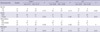
The overexpression of Tspan-1, Jab1 was negatively related to cancer cell differentiation and tumor metastasis (P<0.05). The overexpression of p27 was significantly and positively related to cancer differentiation and tumor metastasis (P<0.05). There was no significantly difference between low expression and over expression of the three proteins in terms of age and gender (P>0.05) (Table 2).
Correlation of the expression of Tspan-1, Jab1 and p27 with the patients' survival
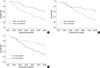
Survival analysis was done in 76 patients in whom follow-up data and results for the expression of Tspan-1, Jab1 and p27 were available. By using the Kaplan-Meier analysis, the patients with Tspan-1 and Jab1 overexprssion are significantly associated with short overall survival (P<0.05) (Fig. 2A, B). Statistical analysis also indicated that the survival rate of the p27 overexpression group was significantly higher than that of the low expression group (P<0.05) (Fig. 2 C). Multivariate analysis using the Cox's proportional hazards model showed that Tspan-1, Jab1 and p27 protein are independent prognostic indicators for patients' overall survival (P=0.018, P=0.012, P=0.002, respectively).
DISCUSSION
Tspan-1, a new member of the tetraspanins group, is a recently discovered tumor-related gene. Some reports had indicated that Tspan-1 can stimulate proliferation, invasion and motility of tumor cells (9, 22). In the present study, we found that the expression of Tspan-1 in HCC tissues was significantly higher than the tissues around cancer or the normal tissues. There is also a strong positive correlation between the level of Tspan-1 expression and degree of HCC cell differentiation and clinical stages. Furthermore, we found that Tspan-1 was an independent factor affecting prognosis of HCC. The over expression of Tspan-1 increased the risk of death. Five-year survival rate in patients with overexpression of Tspan-1 was significantly lower than those patients with low expression of Tspan-1.
In the present study, we found that Jab1 and p27 expression were inversely correlated, and significantly associated with malignancy, prognosis and survival rate in patients with HCC. Similar to our work, other reports indicated that Jab1 overexpression was significantly associated with poor prognosis of patients with tumors such as ovarian tumor, lung cancer and breast cancer, and was inversely associated with p27 (23-25). It has been known that p27 regulate cell proliferation as Cdk inhibitor by inhibiting cell cycle progression from G1 to S phase in a dose-dependent fashion. Jab1 can accelerate the degradation of p27 by translocating p27 from nucleus to cytoplasm where degradation occur (26).
The expression of Tspan-1 in cancer cells displayed cytoplasmic or membranous patterns, which showed the distribution and functional sites of the Tspan-1 molecule in cells. The molecule may accept extracellular signals when located on the membrane and carry out functions in the cytoplasm, like other tetraspanins such as CD9, CD82 and CD63 (27, 28). Leyden et al. reported that siRNA-mediated downregulation of Tspan-1 inhibited the proliferation of gastric cancer cells in vitro by inhibiting cell cycle progression from G1 to S phase (9). In our study, we found that Tspan-1 was positively associated with Jab1, and inversely correlated with p27. It has been known that p27 was degraded specifically in the cytoplasm by the ubiquitin (Ub)-proteasome system (29). So we pursued the question whether Tspan-1 interacted with some other proteins to affect the ubiquitin (Ub)-proteasome system degrading p27 or interacted with Jab1 to accelerate the transportion of p27 from nucleus to cytoplasm, and then led to accelerate the tumor cells proliferation.
In summary, the expression of Tspan-1, Jab1 and p27 in HCC is involved in cancer cell behavior including proliferation, differentiation, metastasis, and clinical stage. Overexpression of Tspan-1 and Jab1 suggested poor prognosis but of p27 may expect good prognosis for patients with HCC.


 XML Download