

 PDF
PDF ePub
ePub Citation
 Print
Print


INTRODUCTION
The Muenke syndrome (MS) is an autosomal dominant craniosynostosis syndrome associated with a mutation in the fibroblast growth factor receptor 3 (FGFR3) gene located on chromosome 4p. The mutation, involving the FGFR3 gene, consists of a heterozygous nucleotide transversion, c.749C>G, encoding the amino acid substitution Pro250Arg. This syndrome is characterized by variable expressivity with unilateral or bilateral coronal craniosynostosis, midface hypoplasia, proptosis, downslanting palpebral fissures, hearing loss, developmental delay, and specific bone anomalies of the hands and feet (1-3).
The frequency of MS has been estimated as 0.8-1.0 in 10,000 live births, with 61% thought to be sporadic cases (4). The mutation rate, at this nucleotide, is one of the most frequently described in the human genome (5). Because of reduced penetrance and variable expressivity with a wide spectrum of clinical findings among patients with MS, in some cases, the typical cephalo-facial anomalies are not present. Therefore, not all family members of affected patients have had molecular evaluation for confirmation of the diagnosis. However, the molecular confirmation of MS is essential for the prediction of risk in affected family members and for the identification of associated anomalies (6).
Here, we report the case of a girl with MS confirmed by molecular diagnosis of the Pro250Arg mutation. Additional family members were identified with this mutation and their clinical characteristics are reported.
CASE REPORT
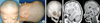
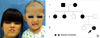
The patient was a 24 month old Korean girl, born at 40 weeks gestation by vaginal delivery at a regional general hospital. The birth weight was 2,900 gm; she was the second child in the family with one female sibling. The mother was 29 yr old and the father was 31 yr old at the time of her birth. The patient showed mild midfacial hypoplasia, hypertelorism, downslanting palpebral fissures, strabismus, a beak shaped nose, and plagio-brachycephaly (Fig. 1A). In addition, the patient had mild neurodevelopmental delay. The hearing was intact and there were no extracranial bony abnormalities. The mother's family had a history of cephalo-facial anomalies. The mother and two of the mother's unmarried sisters and the maternal grandfather had a similar phenotype, strabismus and short stature (Fig. 2). The severity of the brachycephaly was variable among these family members.
Three-dimensional reconstruction computed tomography (3D-CT) and magnetic resonance imaging studies of the cranium revealed bilateral coronal craniosynostosis, palgio-brachycephaly, bilateral lateral ventricle dilatation, proptosis, and a small cerebellum (Fig. 1B, C).
The patient underwent an expansion cranioplasty to correct the brachycephaly at 25 months of age and a second-step surgical procedure was then performed in order to correct the residual posterior plagiocephaly at 5 yr of age.
Mutation analysis
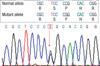
After informed consent was obtained from the parents and other maternal family members, DNA was extracted from the peripheral blood lymphocytes using a standard method with the QIAamp® DNA Blood Mini kit (QIAGEN). The genomic DNA was amplified using the polymerase chain reaction with specific primers. The primer pairs used for FGFR3 exon 7 were forward: GACGTACACGCTGGACGTGCT, and reverse: ACGCAGCTGCCTATGGCCCTGA. The sequences were analyzed by searching for similarities using the BLASTN and BLASTX program at the National Center for Biotechnology Information. The Pro250Arg mutation of exon 7 (IgII-IGIII linker domain) in FGFR3 was detected in the patient with the MS. The same mutation was found in the patient's mother, two maternal aunts and the maternal grandfather (Fig. 3).
DISCUSSION
Craniosynostosis syndromes are associated extracranial anomalies of the limbs, heart, central nervous system (CNS) and/or tracheal malformations. Over 180 different syndromes have been reported in association with craniosynostosis (7). The craniosynostosis syndromes have been reported to be associated with mutations of several genes including: FGFR1, FGFR2, FGFR3, FBN1, TWIST, and MSX2. Among these mutations, the mutations of the fibroblast growth factor receptor gene (FGFR1-3) are the most common and have been implicated in craniosynostosis syndromes (7-10).
The MS is characterized by an extremely variable clinical presentation with unicoronal or bicoronal craniosynostosis, midfacial hypoplasia, ocular hypertelorism, brachydactyly, thimble-like middle phalanges, coned epiphyses, carpal and tarsal fusion, sensorineural hearing loss, and infrequent cognitive impairment (1-3).
The Pro250Arg mutation located between the second and third immunoglobulin-like domains of the FGFR3 gene is commonly found in the autosomal dominant form of craniosynostosis referred to as the MS (11, 12). Different phenotypes have been described in patients carrying the same mutation, Pro250Arg, of the FGFR3 gene (2, 3).
The MS phenotype ranges from no detectable isolated craniosynostosis or macrocephaly to more complex findings that overlap with other classic craniosynostosis syndromes (e.g., Crouzon, Pfeiffer, and Saethre-Chotzen syndromes). Some form of craniosynostosis is involved in most cases of the MS. Bicoronal synostosis is most common; however, some cases have unilateral coronal, lambdoid, and metopic suture synostosis (1-4). A previous report showed that females are more likely to have craniosynostosis with bicoronal synostosis than males (11, 12). Hearing loss has been reported in 25-40% of patients with the MS and a sensorineural impairment is the most common type of hearing loss (65%) (13). The craniofacial phenotype includes facial asymmetry, midface hypoplasia, downslanting palpebral fissures, proptosis, hypertelorism, and a high arched palate (2, 3, 14). No abnormalities of the teeth have been reported. The most common ocular complication is strabismus (3). Extracranial skeletal anomalies occur in 26-42% of patients, including thimble-like middle phalanges, coned epiphysis, carpal and tarsal fusion with fused calcaneocuboid bones, and brachydactyly with broad great toes (11). However, there is no reported case of the MS with associated syndactyly. Developmental delay and learning disabilities have been reported in some of the cases (15, 16). Primary brain anomalies with bilateral dysgenesis of the medial temporal lobe structures have been reported in patients with confirmed MS with typical cranial morphology and molecular testing (17).
Clinical genetic evaluation of patients with the MS should include the following: complete genetic and dysmorphology examination, genetic counseling, referral to a craniofacial team with early intervention, dental assessment for malocclusion, assessment of cognitive function, hearing testing, and ophthalmology assessment. In addition, a newborn hearing screening should be considered in affected families (3).
The corresponding residues of the two other FGFRs involved in craniosynostosis syndromes have mutations; the Pro252Arg mutation of FGFR1 in Pfeiffer syndrome and the Pro253Arg mutation of FGFR2 in Apert syndrome. In addition, the heterozygous Pro250Leu mutation of FGFR3 has been associated with a phenotype similar to MS (12). The Pro250Arg mutation is often familial and associated with a more severe phenotype in females than in males (11, 12). The majority of molecularly defined craniosynostosis syndromes have been shown to have autosomal dominant inheritance. However, unexpected recurrence sometimes occurs due to variable expression; in addition, craniosynostosis can occur in sporadic forms (7, 10). Sporadic cases of the MS have been reported to be associated with advanced paternal age (8).
The patient with MS in this report with the mutation associated with the MS had a phenotype similar to previously reported cases of the MS (Fig. 1). Pedigree analysis demonstrated an autosomal dominant inheritance involving the maternal grandfather, mother and the patient (Fig. 2B). The facial phenotype was similar and strabismus occurred in all affected family members. Short stature was typical and the intelligence was normal. Radiological examination of the skeleton and hearing testing was not performed in the other affected family members.
The cranial sutures are the major sites of bone formation during the embryonic morphogenic period and postnatal skull growth (18). The human FGFRs play an important role in bone and cartilage morphogenesis during the development of vertebrates. Some growth factors (FGFR1-3, and TGFβ) and transcription factors (TWIST, and MSX2) are transiently expressed on the developing surface of the sutures and the ossification regions of primary bones (19-21). Mutations in the FGFR1, FGFR2, FGFR3, FBN1, TWIST, and MSX2 genes have been demonstrated in craniosynostosis syndromes (7-10). More than 89 mutations have been described on the web site of the Human Genome Mutation Database (www.hgmd.cf.ac.uk). The most frequent type of mutation is a missense mutation and the next most common type is splicing errors. Affected patients usually have recurrence in their family members. Premature closure of the cranial sutures, associated with the previously described mutations, can affect the CNS and increase the intracranial pressure. Secondary effects due to CNS involvement include impaired vision, hearing, and severe neurodevelopmental delay (7, 8).
Therefore, molecular confirmation of the associated mutation is important for appropriate genetic counseling and proper patient management. Especially, molecular confirmation of affected family members, so that the correlation between phenotype and genotype can be understood.
This patient illustrates a confirmed Korean case of the MS with autosomal dominant inheritance. The Pro250Arg mutation was confirmed in phenotypically suspicious family members on the maternal side, and the phenotype and genotype correlation in a Koran MS patient were described. Such information is essential for accurate genetic counseling and risk assessment for family members.
 XML Download