

 PDF
PDF Citation
 Print
Print


INTRODUCTION
Otolaryngologic fields (ear, nose, throat, and related structures) are a division of the special senses, and there are impairments of hearing, equilibrium, olfaction, respiration, mastication, deglutition, voice, and speech. Because physical impairments of special senses in otolaryngologic field are subjective, evaluation of physical impairments of special senses in otolaryngologic field is difficult. So we need to make objective standards of physical impairment on the basis of objective clinical data. We accordingly develop a guideline for rating the physical impairment of otolaryngologic fields.
RESULTS
Hearing impairment
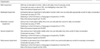
The impairment of hearing disturbance and tinnitus should be assessed by specialists of the otorhinolaryngology in medical institutions equipped with specific instruments. When assessing the impairment, the specialists should first check medical records, certificates and others to show that there is no improvement in the severity of disorders even after a medical treatment of more than 6 months. In the case of possible recovery, the impairment assessment should be held back after medical treatment (1-3).
Required clinical tests are as follows: physical examination, pure tone audiometry, speech audiometry and impedance audiometry. As subsidiary tests, there are brainstem evoked response audiometry (ABR), Bekesy audiometry, otoacustic emission test, and imaging examinations (2, 3).
The results from the pure tone audiometry are the most important in judging the severity of impairment. In addition, other objective methods of audiometry should be supplemented for ensuring the reliability of the test. The pure tone audiometry for hearing disturbance assessment is conducted at the frequencies of 500, 1,000, 2,000, 3,000, 4,000, and 8,000 Hz, and is carried out about three times, once per three to seven days interval (3).
With the pure tone audiogram test results, hearing disturbance is assessed, based on the air conduction pure tone average, according to the six division method (a+2b+2c+d/6, 500 Hz [a], 1,000 Hz [b], 2,000 Hz [c], 4,000 Hz [d]). Not considering the places below the decimal point, in the cases when the auditory threshold in the assigned frequency is above 100 dB or out of the scope of an audiometer, it is regarded to be 100 dB. In the cases when it is below 0 dB, it is regarded to be 0 dB (3, 4).
Having tinnitus with hearing disturbance may lead to the damage of speech discrimination, which can deteriorate the capability of discriminating language. When there is clear and constant tinnitus, which influences the performance of everyday activities and repeated tests show the sound of similar quality and loudness, up to 5% is added to the function impairment (3, 4).
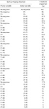
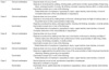
The average hearing acuity of both ears, which is drawn from the test above, is divided into a poorer ear (an ear with worse hearing) and a better ear (an ear with better hearing) in order to assess the impairment. No response indicates an absolute hearing loss, caused by the defective air conduction and bone conduction, and they do not make any response even to the maximum sound pressure of a normal pure tone audiometer. The hearing of above 91 dB means no ability of hearing except for the bone conduction, or the auditory threshold of above 91 dB at the pure tone audiometry. The functional impairment rate is to assess the severity of disorders by the auditory threshold of both ears, by regarding both ears showing no response as 100% hearing loss (Table 1) (4-7).
Balance disorder (Disequilibrium, Vertigo)
Equilibrium sense provides an input to the positions of our own bodies and the sense of direction in space (8). It is maintained by visual system, the proprioceptive system and vestibular organ. As balance disorder can be generated by the disorder of other organs like nervous system, cardiovascular system or visual system, this study deals with the balance disorder produced only by the vestibular disorder (8-10). As the vestibular disorder responds sensitively, the impairment examination should be taken after the illness becomes stable, and the symptoms or signs of impairment should be shown with supportive objective finding (9-11). Furthermore, the examination should consider the functions that are needed in normal activities of examinee. The impairment examination on the equilibrium sense should be taken after making sure by checking medical records and diagnosis that the symptoms continue to be stable even after more than 1 yr of medical treatment by otorhinolaryngologist in specialized medical facilities (8, 9).
History taking, physical examination and radiological examination for the vestibular organ and brain are taken for evaluation. To evaluate the vestibular function, righting reflex test, electronystagmography and calroric test are also taken. When the objectivity of examination is needed, rotatory chair test and posturography can also be used (9-11).
Olfactory disturbance
Olfactory loss or distortion should be evaluated by otolaryngologist with modern means for accurately and objectively assessing olfactory function, including means for detecting malingering (12).
Olfactory perception results from a cascade of events beginning with the arrival of airborne odorant molecules at the olfactory mucosa, and ending in physiological and psychological effects, defining a response to these stimuli. The olfactory receptor cells is a bipolar neuron whose distal process carries cilia, which project into the nasal cavity. These cilia respond to a chemical stimuli by interactions between odorant molecules and receptor proteins on its surface. The proximal nonmyelinated axons form the olfactory nerve, which traverses from the foramina in the cribriform plate to synapse in the olfactory bulb (12).
Anosmia refers to loss of the ability to smell, whereas hyposmia refers to decreased ability to smell. Olfactory dysfunction can be either bilateral or unilateral. Parosmia is distorted or perverted smell perception. Distortion of the sense of smell may bother patients more than the loss of the sense of smell. A problem often encountered in testing olfactory sensitivity is that many patients confuse the loss of the sense of smell with the loss of the sense of taste. Thus, a clear diagnostic distinction should be made between a true taste disorder and an olfactory disorder.
The evaluation of patients with olfactory dysfunction must involve a careful medical history, paying special attention to antecedent events that might be related to the onset of olfactory loss, such as upper respiratory infections, head trauma, nasal surgery, nasal and paranasal sinus disease, and exposure to environmental chemicals.
Essential components of the physical examination include a complete otolaryngologic examination with an emphasis on anterior rhinoscopy and nasal endoscopy, allowing for a thorough assessment of the olfactory cleft. High-resolution computed tomography (HRCT) appears to be the most useful and cost-effective screening tool to assess sinonasal diseases, while magnetic resonance imaging (MRI) is the technique of choice to evaluate the olfactory bulbs, olfactory tracts, and intracranial causes of olfactory dysfunction. In rare instances, biopsies of the olfactory mucosa can be obtained to assess the status of the olfactory epithelium (12, 13).
Olfactory function tests are essential to establish the validity of a patient's complaint, characterize the specific nature of the problem, reliably monitor changes in function over time, detect malingering, and establish compensation for permanent disability. They include University of Pennsylvania Smell Identification Test (UPSIT), Connecticut Chemosensory Clinical Research Center test (CCCRC), T and T olfactometry and Korean Version of Sniffin's Sticks (KVSS) test (12-14).
Despite the fact that a wide range of psychophysical olfactory tests are available for assessing olfactory function, most are of unknown reliability and validity, thus suffering due to lack of normative data. In UPSIT, normosmia scores are over 34, hyposmia scores 18-33, and anosmia scores less than 18. In 1-butanol threshold test, normal subjects score over is 6, hyposmia subjects score 2-5, and anosmia subjects score 1 or 0. In olfactory threshold test by T&T olfactometer, the average recognition threshold is more than 5 in anosmic and 1.1-5 in hyposmic, while less than 1.0 in normal subjects. In KVSS test, Threshold, discrimination, identification (T.D.I.) score is over 31 in normosmic, 15-30 in hyposmic, and less than 15 in anosmic (12, 15).
Malingering sometimes occurs in patients seeking insurance settlements. Malingering is suspected if a patient denies any sensation when the patient is tested with trigeminal stimuli, such as ammonia, acetic acid or menthol. On forced choice psychophysical tests, such as the UPSIT and KVSS, malingering appears with the report of lower scores than expected on the basis of chance (25%) (15, 16).
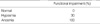
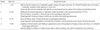
Criteria for evaluating functional impairment in accordance with the degree of olfactory disturbance are those listed in Table 4.
Respiration difficulty
Respiration may be defined as the act or function of breathing, that is, the act by which air is inspired and expired from the lungs. The respiratory system includes the lungs and the air passages; the latter includes the anterior nares, nasal cavities, oral cavity, nasopharynx, oropharynx, hypopharynx, larynx, trachea, and bronchi. Respiratory difficulty can be caused by diseases of the lung parenchyma or defects of the airways. In this proposed guideline, discussion of permanent impairments related to respiration is limited to defects of the air passages (17).
The most commonly encountered defect of air passages is obstruction, which may be partial (stenosis), or complete (occlusion). In patients with airway obstruction, dyspnea is a cardinal symptom that contributes to a patient's diminished capacity to carry out activities of daily living and to permanent impairment. Dyspnea is noted first and is most severe during exercise. However, when dyspnea occurs at rest, respiratory dysfunction is most likely severe. Dyspnea may be accompanied by related symptoms and signs such as voice change, swallowing difficulty, cough, and wheezing.
A complete medical history is important, with specific attention directed toward a history of causative or predisposing disease. Questions about the severity of dyspnea during exercise or at test should be elicited. Other symptoms associated with dyspnea should also be obtained (17-19).
A thorough physical examination is mandatory to evaluate the severity of upper airway obstruction. Chest auscultation may reveal wheezes. The sternal notch and midline neck are examined for evidence of retraction. Obstruction below the thoracic inlet does not cause suprasternal retraction. Endoscopy is the definite diagnostic examination of the upper airway. The examination includes nasopharyngoscopy with a flexible fiberoptic nasopharyngoscope, which is used to assess the airway spanning from the anterior nares to the level of vocal cords. Bronchoscopy can also be performed when the trachea and bronchi are evaluated. Patients with tracheostomy should be evaluated to check whether adequate respiration is possible when the tracheostomy tube is plugged (17).
Anteroposterior and lateral soft-tissue radiography films of the upper airway are often used as screening test for patients with upper airway compromise. HRCT has become an invaluable aid in the evaluation of upper airway, while MRI is very useful for tracheal and laryngeal imaging, which is best performed in the coronal and sagittal planes (17).
The site and character of obstructive airway lesions may be determined by pulmonary function tests with flow-volume loops. Objective measures for the voice may also be needed in patients with abnormal voice.
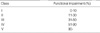
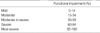
Patients with upper airway defects may be evaluated in accordance with the classification in Table 5 (19).
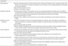
Criteria for evaluating functional impairment in accordance with the degree of airway defects are those listed in Table 6.
Mastication and swallowing difficulty
Mastication and swallowing are essential functions when eating food. A disorder of jaw joint, malocclusion, and tongue illness can cause mastication difficulty. Moreover, esophageal stenosis, tongue illness, and the paralysis of nervous system of pharynx and larynx can cause the swallowing difficulty. When a disorder is generated in the mastication and swallowing function, limitation in eating is inevitable, and thus, becomes the most objective standard to judge the impairment of mastication and swallowing function. Impairment evaluation is taken when the symptoms do not get better and become fixed even with constant treatment of over 1 yr. If doctors foresee improvement in symptoms, reexamination should be taken 2 yr after the final examination. When examining mastication and swallowing function, doctors should see the medical record, operation record, and medical certificate, and should get consultation from dentists if necessary. Evaluation should be taken when the eating ability is stable and the rehabilitation is maximized (20).
The required clinical tests are as follows: history taking, physical examination, examination for upper jaw, lower jaw and temporomandibular joint, dental examination and radiological studies. Endoscopy and esophagography are needed to examine the condition of pharynx and larynx and esophageal obstruction. The scale of impairment is determined according to Table 7.
Voice disorder
Voice disorder refers to an impairment of sound produced by the vocal cord, where there is an abnormality in one of the 3 elements of phonation: intensity (abnormal intensity), pitch (abnormal control), and quality (abnormal quality), which blocks communication. This term is used when objective and medical diagnosis has been made, and the diagnosis of impairment is made only when it is considered permanent after effective treatment of the causative disease. The appropriate timing for the assessment differs depending on the causative disease, and it is done either at the onset or at least 6 months of treatment after surgery. One of the exception to this rule is total laryngectomy, where the diagnosis can be made immediately after surgery. Vocal function can be divided into near-distance vocal function and daily vocal function. The near-distance function is the ability to communicate with family members or the care-giver to carry out basic daily life and can be assessed by having patient read a few sentences and asking a few questions within 1.5 m distance. Daily life vocal function is the ability to adequately manage and communicate in the vocational-social life.
Voice disorder is easier to diagnosis and is more objective when using the guidelines based on anatomical loss or derangements, but it does not necessarily correlate with the actual function. However when using the guidelines based on the function, it has limitations in that it requires the examinees to actively participate, and such cooperation is inevitable. The same diagnosis holds many levels of impairment and it is difficult to find an objective means of the rating (21).
Currently, there is no single objective method available to measure the rate of voice disorder, therefore, we measure it with various methods. The compulsory tests used include physical, oral, pharynx, and larynx endoscopic examination, larynx stroboscopy, and hearing assessment by a speech-language pathologist, and some of the supplementary tests include laryngeal electromyography, computerized sound analysis test, aerodynamic test, electorglottography, and radiologic examinations (CT and MRI) (Table 8, 9).
Articulation impairment
Articulation means using organs of phonation to communicate with others in everyday sense, therefore, articulation difficulty refers to the limitation of communication using spoken language. It is limited to the cases in which objective and medical methods have been used in diagnosis, and in cases of aphasia due to the destruction of language center in the central nervous system, and language development disorder in developing age eliminated. Thus, articulation difficulty in broad terms can be made by consulting neurology, rehabilitative medicine, pediatrics, and psychology department. Rating of voice disorder and articulation difficulty should be done separately, and the higher degree of impairment between the two is used as a principle.
Various objective methods are used for assessment, and every assessment should be made by a speech language pathologist. In clinical sense, fluency disorder can be assessed by paradise-fluency assessment (P-FA) and stuttering severity instrument (SSI). Articulation disorder is assessed by Picture consonant articulation test (PCAT). There are position articulation test, Lee-Kim Korean articulation picture (KAP), and speech intelligibility assessment.
In making the diagnosis of articulation disorder, language analysis should be made on the patient's major speech problems. In such a case, pronunciation test should be done to assess the consonant accuracy. If it is above 76%, speech intelligibility test should be made to rate the impairment (Table 10, 11).
DISCUSSION
In impairment of hearing disturbance and tinnitus, previous guideline for rating the hearing impairment (the State Tort Liability Act) was made based on condition of tympanic membrane and subjective hearing ability according to distance. Previous guideline divided hearing impairment into six degrees (20%, 30%, 40%, 60%, 70%, and 90%). We developed more objective guideline for hearing disturbance based on pure tone audiometer. We can rate hearing impairment precisely by checking hearing ability of better and worse ear.
In impairment of balance, previous guideline for rating the balance impairment (the State Tort Liability Act) was made based on degree of working disability due to disturbance of neurologic function. Previous guideline divided balance impairment into two degrees (40% and 60%). We developed more objective guideline for balance impairment based on laboratory findings (righting reflex test, electronystagmography, calroric test, rotatory chair test, and posturography), treatment history and functional impairment findings.
In impairment of olfaction and respiration, previous guideline for rating the olfactory and respiratory impairment (the State Tort Liability Act) was made based on physical finding such as nasal deformity and degree of neurologic symptom. Previous guideline divided olfactory and respiratory impairment into three degrees (5%, 15%, and 40%). We developed more objective guideline for olfactory and respiratory impairment based on clinical manifestation and laboratory findings (UPSIT, CCCRC, KVSS, flexible fiberoptic nasopharyngoscope, bronchoscopy, simple soft-tissue radiography films of upper airway and HRCT). In new guideline, we considered anatomical state of upper and lower respiratory tract. So we can more precisely rate respiratory impairment.
In impairment of mastication and swallowing, previous guideline for rating the mastication and swallowing impairment (the State Tort Liability Act) was made based on subjective mastication and swallowing function of patient. Previous guideline divided olfactory and respiratory impairment into seven degrees (5%, 15%, 30%, 40%, 70%, 90%, and 100%). We developed more objective guideline for mastication and swallowing impairment based on history taking, physical examination, examination for upper jaw, lower jaw and temporomandibular joint, dental examination and radiological studies. Endoscopy and esophagography are used to examine the condition of pharynx and larynx and esophageal obstruction. In new guideline, we considered anatomical state of digestive tract. So we can more precisely rate mastication and swallowing impairment.
In impairment of voice, previous guideline for rating the voice impairment (the State Tort Liability Act) was made based on subjective phonation function of patient. Previous guideline divided voice impairment into seven degrees (5%, 15%, 30%, 40%, 70%, 90%, and 100%). We developed more objective guideline for voice impairment based on history taking, oral, pharynx, and larynx endoscopic examination, larynx stroboscopy, hearing assessment. We can use supplementary tests such as laryngeal electromyography, computerized sound analysis test, aerodynamic test, electroglottography, and radiologic test (CT and MRI). In new guideline, we considered anatomical state of phonation system. So we can more precisely rate voice impairment.
In impairment of articulation, previous guideline for rating the articulation impairment (the State Tort Liability Act) was made based on subjective articulation function of patient. Previous guideline divided articulation impairment into seven degrees (5%, 15%, 30%, 40%, 70%, 90%, and 100%). We developed more objective guideline for articulation impairment based on history taking, physical examination, fluency test by P-FA and SSI, articulation test by PCAT, position articulation test, KAP, and speech intelligibility assessment. We consider consonant accuracy and stuttering when we rate articulation impairment. In the new guideline, we consider anatomical and functional state of articulation system. So we can more precisely rate mastication and articulation impairment.

 XML Download