

 PDF
PDF ePub
ePub Citation
 Print
Print


INTRODUCTION
Drug-induced neutropenia (DIN) is a rare, sporadic, and transient disorder. The patients with DIN experience a high rate of infectious complications and have a mortality rate of approximately 10% (1, 2). The most common drugs associated with DIN are antithyroidal drugs, anticonvulsants, and antibiotics such as cephalosporins, penicillins, sulfonamides, and chloramphenicol, although the pathogenesis of DIN is poorly understood (3).
Here, we describe the brief history of a child with pneumococcal pneumonia, who experienced severe neutropenia during antibiotic treatment and in whom we detected 4 kinds of antibiotic-dependent antineutrophil antibodies.
CASE REPORT
A 22-month-old boy presented to another hospital with fever and cough for 3 days. On admission to our institution, he was irritable and dyspneic. Physical examinations revealed markedly decreased breathing sounds over the left lung fields. The radiologic findings showed diffuse infiltration in the left lower lung fields without shifting of pleural fluid. Laboratory studies on admission disclosed the following values: hemoglobin 11.2 gm/dL, white blood cell (WBC) 9,800/µL (differential count; segmented neutrophil 82.6%, lymphocyte 14.8%, monocyte 2.6%), platelet 281,000/µL, erythrocyte sedimentation rate (ESR) 55 mm/hr, C-reactive protein (CRP) 32.2 mg/dL. We started antibiotics with augmentin (100 mg/kg/day, intravenously) and tobramycin (5 mg/kg/day, intravenously) as well as intravenous immunoglobulin (IVIG, 1 gm/kg) after septic workup including blood cultures. Three days later, we switched antibiotics to vancomycin (50 mg/kg/day, intravenously), cefotaxim (200 mg/kg/day, intravenously), and roxithromycin (8 mg/kg/day, orally) because the blood culture revealed Streptococcus pneumoniae being sensitive to vancomycin and the antibody titer for Mycoplasma pneumoniae was 1:160 even with a negative cold agglutinin test. His clinical condition became well without fever after 8 days of antibiotic therapy.
On hospital day 12, before which it was not evident, fluid shifting on a chest radiography was detected and also confirmed by chest computed tomograohy (CT) scans. A chest tube was inserted and pleural fluid analysis showed compatible findings with transudate, possibly due to previous antibiotic therapy. Furthermore, polymerase chain reactions for Mycobacterium tuberculosis and Mycoplasma pneumoniae in pleural fluid were also negative. The serologic test for Mycoplasma pneumoniae antibody was 1:160 and cold agglutinin test was negative. The blood tests disclosed hemoglobin 6.2 g/dL, WBC 18,500/µL (differential count; segmented neutrophil 62%, lymphocyte 35%, monocyte 3%), platelet 979,000/µL, reticulocyte 2.3%, ESR 68 mm/hr, CRP 4.32 mg/dL. He did not receive packed red blood cells (RBC) because of a positive direct Coombs test and warm antibody on the screening test. He received aspirin because of a high platelet count.
On hospital day 15, fever developed again and persisted until hospital day 18, when progressive, generalized, erythematous maculopapular rashes also appeared and a complete blood count (CBC) revealed hemoglobin 7.8 g/dL, WBC 4,200/µL (differential count; segmented neutrophil 29%, lymphocyte 59%, monocyte 9%, eosinophil 1%, atypical lymphocyte 1%, myelocyte 1%), platelet 224,000/µL, and reticulocyte 7.3%. Generalized skin rashes and edema were aggravated until hospital day 22, when IVIG and solumedrol was administered with replacement of vancomycin by same doses of augmentin and tobramycin after removal of the chest tube. Thereafter, the fever and skin rashes disappeared, even the CBC findings on hospital day 26 showed severe neutropenia (absolute neutrophil count: 492/µL).
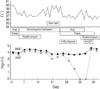
On hospital day 32, the fever developed again without any significant symptoms or signs. CBC findings on next day disclosed severe neutropenia with WBC 3,700/µL (differential count; segmented neutrophil 1%, lymphocyte 91%, monocyte 8%). Severe neutropenia persisted until withdrawal of augmentin and tobramycin as well as administration of granulocyte colony-stimulating factor (G-CSF, 5 mcg/kg/day, subcutaneously) for 3 days from hospital day 36 when the blood samples for antineutrophil antibodies were collected. The brief clinical course as well as the changing pattern of absolute neutrophil counts is presented in Fig. 1.
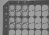
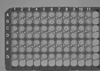
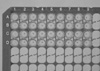
To detect neutrophil antibody, we used the mixed passive hemagglutination assay (MPHA), using extracted neutrophil antigens coated onto microplates (4-6). Neutrophil antibody IgG was detected in the patient's serum. The serum was reactive with the patient's neutrophil but not with donors' neutrophil (Fig. 2). Furthermore, positive control sera were reactive with all donors' neutrophil because human neutrophil antigen (NA 1 and NA 2) are present in the all donors' neutrophil including patient's neutrophil, but patient's own antibodies did not have specificity for NA 1 and NA 2. The serum was 1:8 positive with the patient's neutrophil. The serum incubated with cefotaxim, augmentin, vancomycin, and tobramycin was positive (all 1:64) with the patient's neutrophil, but the serum incubated with aspirin and roxithromycin was less reactive (1:4) with the patient's neutrophil than the intact serum (Fig. 3). The serum coincubated with drugs was not reactive with any of the donor's neutrophils (Fig. 4). In conclusion, there was an anti-neutrophil autoantibody that had specificity for antibiotics (cefotaxim, augmentin, vancomycin, and tobramycin).
DISCUSSION
DIN is an idiosyncratic reaction that is mediated by immune or allergic and toxic mechanisms, and results in profound neutropenia (7). Although antineutrophil antibodies also appear in a range of diseases associated with neutropenia, such as pure white blood cell aplasia, immune neutropenia, Felty's syndrome, and systemic lupus erythematosus, antineutrophil antibodies have been detected in most of the cases of antibodies to autologous or normal granulocytes in immune-mediated DIN (8). In DIN, antibody binding to a target cell usually requires the presence of the causative drug and the complement is often consumed (9). Other mechanisms, involving cytotoxic T cells, haptens, autoimmunity and oxidative modifications of drugs as well as direct damage to myeloid precursors have been reported (10-13).
The causes of neutropenia include infectious diseases (especially viral), exposure to chemicals, nutritional deficiencies, immune disorders, and congenital or chronic neutropenia (7). However, most, but not all, instances of neutropenia result from exposure to drugs, and either the drug itself or a metabolite may be causative (3). The annual incidence of symptomatic nonchemotherapy DIN is between 2.4 and 15.4 cases per million people per year (7). This incidence increases with age, as only 10% of cases are reported in children and young adults and more than half of these episodes occur in people aged over 60 yr (14).
The drugs most commonly associated with neutropenia are antibiotics (particularly beta-lactam and trimethoprim-sulfamethoxasole) as well as antithyroid drugs, antiplatelet agents, nonsteroidal anti-inflammatory agents, and noramidopyrin (3). We experienced a case of DIN in a child in whom was detected antineutrophil antibodes by MPHA technique to 4 kinds of antibiotics, but not to roxithromycin and acetylsalicylic acid. Even if it is not usual to detect antibodies simultaneously for 4 kinds of antibiotics, we could not try to confirm the basic mechanism with further studies. In this child, fever and allergic skin reactions developed respectively at 17 days, 10 days prior to neutropenia, which suggested a vancomycin induced drug reaction. Our patient also showed low hemoglobin level at 20 days prior to detection of severe neutropenia, which could be considered to be immune mediated hemolytic anemia. However, we could not find laboratory evidence of hemolysis except from the positive direct Coombs test, which was considered to be the effect of previous administration of intravenous immunoglobulin. We believed that the decreased hemoglobin level was caused by the suppression of bone marrow function due to severe infection, although we did not perform bone marrow examinations, and eventually the decreased hemoglobin level was normalized without transfusion in accordance with the control of infection. In conclusion, we could suggest that neutropenia occurred in this patient was mediated by autoantibodies to antibiotics, based on the immune mediated clinical findings which showed prior to detection of antineutrophil antibodies in patient's serum. Thus we report this patient with DIN presenting antineutrophil antibodies to antibiotics.
 XML Download