

 PDF
PDF ePub
ePub Citation
 Print
Print


INTRODUCTION
Endothelin has been identified as a neurohormone that is activated during heart failure, and also by renin-angiotensin-aldosterone, epinephrine-norepinephrine, and natriuretic peptide. In addition, a number of studies have shown that endothelin-1 and its receptors over ischemic myocardial tissue are increased during ischemia-reperfusion injury. The endothelin-1 is the principal isoform in disease states and also is the most potent vasoconstrictor. It induces myocardial injury during ischemia and reperfusion as a contributor (1).
There are numerous reports about the relationship between endothelin and congestive heart failures (CHF). Some authors showed that endothelin-1 is an independent, noninvasive predictor of functional and hemodynamic response to therapy in patients with CHF. The majority of heart failure is caused by cardiomyopathy, and the increased endothelin levels have also been observed in the cardiomyopathy heart such as cardiomyopathic Syrian hamsters (2) or in the experimental Chagas' cardiomyopathic hearts infected with Trypanosoma cruzi (3).
Tezosentan, the first intravenous endothelin A and B receptor antagonist, has been evaluated as the treatment for acute and chronic heart failure. There are two large randomized controlled studies about tezosentan on acute heart failure syndrome-RITZ (randomized intravenous tezosentan study) and VERITAS (value of endothelin receptor inhibition with tezosentan in acute heart failure studies) (4, 5). Although these studies demonstrated mixed results of tezosentan as the drug of treatment for decompensated heart failure, the roles of the myocardial protective effect of tezosentan in various heart failure models have not yet been defined. The objectives of this experiment are to evaluate the myocardial protective effects of tezosentan in various experimental heart failure models.
MATERIALS AND METHODS
Animals
Total 90 Sprague-Dawley male rats (6-8 weeks old, 200-300 g) were housed at 20±2℃ and 55±20% humidity with 12-hr light/dark cycles, and free access to food and water in the Animal Care Facility at the Ansan Hospital, Korea University, Korea. This study was approved by the Committee of Animal care of Korea University and conforms with the Guide for the Care and Use of Laboratory Animals published by the US National Institutes of Health (NIH Publication No. 85-23, revised 1996).
Ischemic cardiomyopathy model (ICMP)
Rats were anesthetized with ketamine (100 mg/kg) andxylazine (10 mg/kg), intraperitoneally. The endotracheal intubation was performed, and positive pressure ventilation was given using a mechanical ventilator (Harvard Apparatus, South Natick, MA, U.S.A.) with room air at a respiratory rate of 55-65 strokes/min and tidal volume of 10 mL/kg body weight to maintain normal PO2, PCO2 and pH of blood.
Under the right lateral decubitus position, the left anterior descending coronary artery was ligated with a 6-0 silk suture through left anterior thoracotomy. The lungs were then inflated and the chest incision was closed. After recovery of self respiration, extubation was performed.
Doxorubicin-induced cardiomyopathy model (DOX)
Doxorubicin hydrochloride (D1515, Sigma Chemical Co, St. Louis, MO, U.S.A.) was administrated in 6 equal injections (each intraperitoneal injection containing 2.5 mg/kg in saline, for a total dose of 15 mg/kg) during 2 weeks. Rats were observed for their general appearance, behavior, and mortality for 4 weeks after the final injection.
Pressure-overload hypertrophy model by transverse aortic constriction (TAC)
Under sterile condition, the midline cervical incision was performed; 1-2 cm incision was made at the level of cricoid process and dissection down to the clavicle. After cutting of the right clavicle, the thymus was retracted gently, and the aortic arch was exposed. A constriction of the aortic arch was conducted between the both carotid arteries using a 16 gauze angiocatheter (internal diameter 0.6 mm).
Experimental design
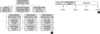
We divided the experimental animals into three cardiomyopathy groups; ischemic dilated cardiomyopathy group (ICMP, n=30), doxorubicin-induced cardiomyopathy group (DOX, n=30) and pressure-overload hypertrophy group by transverse aortic constriction method (TAC, n=30). One month after the construction of experimental rat groups, each group was further randomly subdivided to a sham-operated ischemia-reperfusion subgroup (SHAM) operation (SHAM, n=10), tezosentan treatment ischemia-reperfusion subgroup (Tezo, n=10), or tezosentan non-treatment ischemia-reperfusion subgroup (N-Tezo, n=10) (Fig. 1A). In the SHAM subgroup, only open and closure procedures or injection of saline instead of doxorubicin were performed.
Ischemia-reperfusion protocols
Four weeks after the construction of cardiomyopathy models, the rats were anesthetized with sodium pentobarbital (50 mg/kg, intraperitoneally). Hearts were rapidly excised, connected via the aorta to Langendorff apparatus (size 3, type 830, Hugo Sachs Elektronik, March-Hugstetten, Germany) and perfused retrogradely at a constant pressure of 80 mmHg with the perfusate (Krebs-Henseleit solution) of the following composition: NaCl, 118.1 mM; KCl, 4.6 mM; CaCl2, 2.5 mM; MgSO4, 1.2 mM; KH2PO4, 1.2 mM; NaHCO3, 24.8 mM; and glucose, 10 mM. The perfusate was continuously bubbled with a gas mixture of 95% O2/5% CO2 (pH 7.4), and temperature was maintained at 37℃ throughout the experiment. A latex balloon filled with water was inserted into the left ventricle through the left atrium and attached to a pressure transducer (DX-360; Nihon Kohden, Tokyo, Japan). After 30 min of stabilization period, total circulatory arrest was performed with cardioplegia and coldness about 1 hr. After then, reperfusion was continued for 2 hr. In Tezo subgroup, tezosentan 10-5 M/L was added to the cardioplegic solution and the perfusate (Fig. 1B), but not to N-Tezo subgroup.
Drugs
Tezosentan (Ro 47-0203, Actelion Pharmaceuticals Ltd, Allschwil, Switzerland) is a non-selective specially formulated intravenous dual (endothelin-A and -B) endothelin receptor antagonist. Unlike its sister compound (bosentan), tezosentan is easily soluble in water and stable in solution at physiologic pH, therefore, usable clinically. Another drug, doxorubicin hydrochloride (D1515, Sigma Chemical Co) is well known for cardioselective toxicity and induces congestive heart failure.
Hemodynamic studies for cardiac performance
The left ventricular pressure was monitored with a pressure transducer to obtain the peak positive and negative first derivatives (dP/dTmax and dP/dTmin), and the left ventricular developed pressure (LVDP) was calculated as the difference between the left ventricular (LV) systolic and diastolic pressures. These parameters were measured by an amplifier (AP601G; Nihon Kohden) and also recorded using PowerLab/4sp software (AD Instruments, Mountain View, CA, U.S.A.). Coronary flow was also determined by collecting the coronary effluent from the hearts.
RESULTS
Left ventricular developed pressure (LVDP)
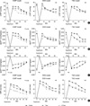
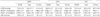
LVDP of Tezo subgroup was significantly higher than that of N-Tezo subgroup in the ICMP group after 30 min of reperfusion (64.3±7.1 mmHg vs. 41.4±4.3 mmHg; P<0.05). In the DOX or TAC model, LVDPs were not significantly different between Tezo and N-Tezo subgroup during the entire reperfusion period (Fig. 2A, Table 1).
dP/dTmax
In the ICMP group, dP/dTmax was significantly different among the three subgroups at 30 min of reperfusion (SHAM, 3,491±21 mmHg/sec; Tezo, 2,821±13 mmHg/sec; N-Tezo, 2,282±182 mmHg/sec; P<0.05). In the DOX or TAC groups, there were no statistical differences among three subgroups (Fig. 2B, Table 1).
dP/dTmin
In the ICMP group, dP/dTmin was significantly different among the three subgroups at 30 min of reperfusion (SHAM, -2,382±114 mmHg/sec; Tezo, -1,638±130 mm Hg/sec; N-Tezo, -1,387±81; P<0.05), similar to the above dP/dTmax. In the DOX or TAC groups, there were no statistically significant differences among the three subgroups (Fig. 2C, Table 1).
DISCUSSION
The effect of endothelin-1, the principal isoform of endothelin, is mediated by 2 types of receptor, endothelin-A and endothelin-B. The endothelin-A receptor appears to mediate vasoconstriction and stimulate the secretion of atrial natriuretic peptide, while the endothelin-B receptor mediates endothelin-induced vasodilatation and activation of the renin-angiotensin-aldosterone system. Through these effects, endothelin has been demonstrated to be not only one of the most potent vasoconstrictors known, but also to mediate pathologic hypertrophy and fibrosis of both ventricular and vascular tissues, thus promoting progression of atherosclerosis, ventricular and vascular remodeling (6-8).
Tezosentan, a dual inhibitor of endothelin A and B receptors, is weakly acidic, highly soluble in water and stable in physiologic pH solution. These properties make it an ideal candidate for intravenous use, and are the reason of why we used tezosentan in our study.
Heart failure has been associated with elevated plasma levels of endothelin-1, and the magnitude of the elevation with the severity of disease, the incidence of arrhythmia, and a worse prognosis. Increased endothelin levels have been described in patients with acute and chronic CHF that are predictive of increased mortality risk (9). Furthermore, increased endothelin levels have been suggested to play an important role in the increased systemic vascular resistance seen in CHF (10). Therefore, endothelin seems to play a major role in the pathogenesis of experimental and clinical heart failure (11, 12).
Although the endothelin receptor antagonists may have the ability to improve the outcome of heart failure by reducing preload and afterload, increasing the contractility of failing myocardium, delaying myocyte hypertrophy, and reducing the incidence of arrhythmias, some studies demonstrated no benefit of the endothelin receptor antagonists for myocardial ischemia-reperfusion heart models (13-15). Therefore, we aimed to investigate the myocardial protective effects of tezosentan for the ischemia-reperfusion injury in various heart failure models.
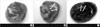
Before analyzing our experimental data, it was necessary to confirm the in-depth of animal heart failure models. Therefore, one month after cardiomyopathic animal models had been constructed, they were verified with pathologic specimens during euthanasia of the experimental animals. In the ICMP models, staining of triphenyltetrazolium chloride with evans blue was used for visualizing ischemia-infarct area of myocardium. In the DOX models, dilated myocardium was confirmed with pathologic specimens and hypertrophied myocardium was also confirmed in the TAC models (Fig. 3).
We also believe that hemodynamic parameters such as LVDP, dP/dTmax, dP/dTmin and CBF are adequate in our present study to verify myocardial protective effect of tezosentan in various rat cardiomyopathy models. It should be mentioned that these parameters have been used for proving drug efficacy in Langendorff technique (16).
As a result, our present study did not reveal any significant effect of tezosentan on DOX and TAC models. There are some hypotheses and limitations on this result in our study. First, although heart failure has been associated with elevated plasma levels of endothelin-1, the density of endothelin and endothelin binding sites had been produced in the heart has not been uniform among different animal heart failure models (17). In myocardial infarct model, it is well established that the infarcted myocardium induces the myocardial necrosis, and increases endothelin-1 levels many fold not only in the post-acute myocardial infarct scar tissues but also in the healed infarct scars after 4 and 13 weeks (18-20). Therefore, endothelin-1, a recognized fibrogenic factor, plays a crucial role in the stabilization of the necrotic area and in the healing of the scar (20). However, the most prominent histological feature of DOX is not the myocyte necrosis, but the loss of myofibrils and cytoplasmic vacuolization caused by dilatation of the sarcoplasmic reticulum in the myocardial cells (21), and there is a report that high dose of endothelin antagonist is needed to block endothelin induced hypertension in the dilated cardiomyopathy rat models (22). In addition, sustained pressure overload stimulate pathological cardiac hypertrophy and dysfunction (23) and hypertrophy progresses to decompensated phase with cardiac dilatation and contractile impairment during chronic pressure overload (24, 25). Some authors showed that the production of endothelin-1 increased in the hypertrophied left ventricle (26), but it depends on the time of the disease progress. Ventricular endothelin-1 levels and the density of endothelin-1 binding sites were increased significantly, without affecting their binding affinity, on day 8 of pressure overload (27), after then, the production of endothelin-1 is decreased and endothelin receptors are down-regulated in hypertrophied ventricles 8 weeks after aortic coarctation (28). However, it is also reported that endothelin-1 levels in subjects with heart failure in whom they are found to be elevated in the late, decompensated phase of the disease (29). This time-dependant up- and down-regulations of endothelin and endothelin receptors are also another proof for our hypothesis. That is, because of the differences of pathophysiology among our study group, cardiac endothelin-1 and endothelin receptors might not uniformly be induced in various heart failure models.
Second, in addition to endothelin, renin-angiotensin-aldosterone, epinephrine-norepinephrine, and natriuretic peptide have also been identified as neurohormones to be activated in cardiomyopathy (30). It is well known that doxorubicin administration has been shown to activate the renin-angiotensin system (31, 32) and the atrial natriuretic peptide level has also been increased relative to the severity of the cardiac dysfunction in human (32). And, there is a study on angiotensin and angiotensin receptor antagonist in the hypertrophic cardiomyopathy (33), showing that stimulation of the angiotensin receptor has an important role in disease process during pressure overload. Therefore, these neurohormones might be more important disease mediators in the DOX or TAC models than in the ICMP model.
There was a limitation to our study. Endothelin is a stimulator of polymorphonuclear leukocyte (PMN) aggregation (34) and adhesion as well as a potent vasoconstrictor. Myocardial ischemia initiates an acute inflammatory response in which PMNs are of importance (35). Because we had used isolated heart perfusion using Langendorff technique, however, most of the PMNs were diluted in the buffer perfused isolated heart. In other words, the effect of PMNs might be restricted and it can affect the results in our study.
In conclusion, our result reveals that tezosentan has myocardial protective effects for the ischemia-reperfusion injury in the ischemic cardiomyopathy model, but not in the doxorubicin-induced cardiomyopathy and pressure-overload hypertrophic heart failure models.
 XML Download