

 PDF
PDF ePub
ePub Citation
 Print
Print


INTRODUCTION
Although the incidence of bacterial pneumonia has steadily decreased according to the improved public hygiene in the developed countries, Streptococcus pneumoniae is still one of the most common pathogens of childhood empyema (1). Transient hypogammaglobulinemia of infancy (THI) is originally defined as a physiological maturation defect of immunoglobulin G (IgG) production that occurs at 3-6 months of age and it lasts until 18 to 36 months of age. The majority of children with THI may be asymptomatic, but children with recurrent infections have been incidentally detected as having THI (2-5). We report here on a 22-month-old child with THI and IgA deficiency and she had massive pneumococcal empyema. After this episode, she has not experienced recurrent infections or a severe infectious episode, and she has shown a normal growth pattern. Her IgG level returned to normal within 6 months, but IgA level is still low at 6 yr of age.
CASE REPORT
A 22-month-old girl was admitted to our hospital because of dyspnea and peripheral cyanosis for 2 days. She was born at full term and had been healthy until this event. She had no known history of severe infections and no familial history of immunodeficiency. Before admission, she had complained of cough with sputum for 2 weeks and she had visited private clinics 3 times. Fever was noticed for 2 days at the first visit to the clinic, since then she had remained afebrile. The weight, height and head circumference of the patient were within the normal percentile ranges for her age. Laboratory investigations revealed hemoglobin 14.9 g/dL, white blood cell count 14,000/µL (66% neutrophils and 30% lymphocytes), platelet count 123,000/µL, erythrocyte sedimentation rate at 1 hr 2 mm/hr and C-reactive protein 0.1 mg/dL. The blood chemistry analysis was non-specific except elevated alkaline phosphatase 685 IU/L (96-254 IU/L) and lactate dehydrogenase (LDH) 823 IU/L (145-420 IU/L). The serum complement levels were C3 61.4 mg/dL (77-195 mg/dL), and C4 10.0 g/dL (7-40 mg/dL).
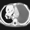
A chest computed tomography (CT) performed on admission day showed massive pleural effusion with a totally collapsed left lung, and the heart was shifted to the right side (Fig. 1). The values of immunoglobulins on the 14th admission day were IgG 336 mg/dL (reference level for age: 345-1,236 mg/dL), IgA <13 mg/dL (14-159 mg/dL), IgM 87.6 mg/dL (43-207 mg/dL) and IgE 31 IU/mL (0-170 IU/mL). Although all the values of IgG subclasses were low, there was no IgG subclass that was not detected. Isohemagglutinin and the antibodies from vaccination (anti-diphtheria IgG, anti-tetanus IgG and anti-polio virus IgGs) were all detected. The lymphocyte subset tests showed that the pan-T cells were 51.6% (28-77%), the CD4+ cells 25.3% (32-68%), the CD8+ cells 23.6% (10-36%) and the B cells 36.5% (10-20%). The nitroblue tetrazolium test was negative. The degree of T cell proliferation to mitogens (phytohemagglutinin and anti-CD3/anti-CD 28 monoclonal antibodies) was comparable to that of the age-matched control.
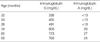
On the second day of hospitalization, a chest tube was inserted to the pleural cavity and ~300 mL of milky colored fluid was evacuated. The pleural fluid analysis revealed an exudate with subsequent heavy growth of S. pneumoniae; the organism was resistant to penicillin, but sensitive to ceftriaxone. The clinical course of the patient treated with the chest tube drainage and an antibiotic regimen (ceftriaxone) was uneventful and there was no residual lesion on the chest radiograph taken 6 months later. The patient did not receive intravenous immunoglobulin therapy during hospitalization or subsequent supplementary therapy for hypogammaglobulinemia. After 1 yr, the low levels of IgG and C3 and the ratio of CD4+ T cells were recovered to within the normal range for her age, but her IgA level did not recover (Table 1).
DISCUSSION
The immune system of early childhood is not fully matured compared to adults, and some immune functions may mature during childhood. The phenotype of some infectious diseases such as hepatitis A is age-dependent, and most children under 5 yr of age are largely asymptomatic or innocuous without jaundice when they are affected by hepatitis A virus (6). Kawasaki disease is an acute febrile systemic vasculitis of an unknown etiology, but it may be an immunemediated disease that manifests after an infection by unknown pathogens (7). This disease has a strict age predilection between 6 months and 4 yr of age. In addition, other early childhood immune-mediated disorders that have a strict age restrictions with a self-limited clinical nature are known; transient erythroblastopenia of childhood (TEC) (8), autoimmune neutropenia of infancy (ANI) (9), and childhood immune thrombocytopenic purpura (ITP) (10). All these findings indicate that the maturing immune system in early childhood (< 5 yr of age) may be involved in the phenotype of infectious and immune-mediated disorders. THI also occurs mainly at 3-6 months of age and it lasts until 18 to 36 months of age with self-limiting recovery. Thus, it may be regarded as one of the disease category that reflects immunologic immaturity during early childhood. The pathogenesis of THI remains unknown, although delayed functional maturation of B cells, a numerical reduction of helper T cells (also suggested in our case), and cytokine imbalance have been postulated (2-5, 11, 12). Most of the children with TEC, ANI or ITP are proposed to experience a variety of infections, and especially those of a viral origin before presentation of illness. It is postulated that THI may also be associated with an infection and the subsequent immune perturbation is responsible for the pathogenesis of THI.
Neutrophils and other phagocytes are the first defense line against extracellular bacterial pathogens, and humoral immunity (antibodies) has a crucial role for phagocytosis against encapsulated bacteria such as S. pneumoniae and H. influenzae. Thus, the patients with humoral immunodeficiencies such as X-linked agammaglobulinemia (XLA) and common variable immunodeficiency (CVID) mainly complain of recurrent infections from such bacteria (13).
A majority of children with THI are detected by clinical manifestations like recurrent upper respiratory infections, but they have few severe infections during the follow-up period (2-5). Some children with THI experience severe or life-threatening infections such as sepsis or severe pneumonia, like happened in our case (14-16). Since the patients with THI are believed to have a normal capacity to produce specific antibodies, in contrast to XLA and CVID patients, the low level of IgG alone may not be responsible for a severe infection with encapsulated bacteria. Our patient was also noted to have an intact humoral immunity and a normal T cell proliferation response with a decreased CD4+ T cell count. The low levels of IgG, C3 and CD4+ T cells were recovered within 6-12 months. Thus, other transient immune disturbances concerned with phagocytosis or other immune function may manifest severe infections in some of the patients with THI, including our case, although we did not perform extended immunologic studies at presentation.
Previous studies have indicated that a subgroup of THI patients had a persistent low value of IgG beyond >4 yr of age and this was commonly associated with low IgA values (IgA immunodeficiency) or dysgammagrobulinemia (2-5, 14). These findings suggest that THI may consist of a heterogenous group of immunodeficiencies and THI may have the possibility of an early manifestation of a primary immunodeficiency such as CVID, although there are few reports on the association between THI and primary immunodeficiency.
Although THI is an age-dependent and self-limiting disorder, severe infections may occur, including atypical presentation of infections that need immunologic study. Long-term follow-up of the clinical condition of these patients and their immunologic status are necessary to rule out primary immunodeficiency disorders.
 XML Download