

 PDF
PDF ePub
ePub Citation
 Print
Print


INTRODUCTION
Open fracture of distal femur with popliteal artery occlusion could be a challenge to many orthopedic surgeons (1, 2). Moreover treatment of severe bone loss of distal femur would also be considerably more difficult because of complete loss of ligamentous stability of knee joint, and involvement of disruption to the knee extensor mechanism (3, 4).
In this article, we report a case of severe bone loss of distal femur with popliteal artery occlusion. After revascularization of the limb, we successfully performed a reconstructive total knee arthroplasty with modular segmental endoprosthesis, and augmentation of patellar tendon using a semitendinosus allograft.
CASE REPORT
Paramedics brought a 45-yr-old man into our medical center immediately after a high-velocity crush injury. On admission to the emergency room, the patient was noted to have a large open wound in anterior aspect of left knee. His left foot was pale and cool. Distal pulses of dorsalis pedis artery and posterior tibial artery were absent, and only femoral pulse was palpable on the injured left lower extremity. He was alert with respiratory rate of 22, pulse rate of 98, and blood pressure of 100/80. Neurological examination relating to his left leg was grossly normal.
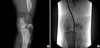
The knee joint was open, and a portion of the patellar tendon was lost. About 50% of the patellar tendon was remained, but it was detached from the inferior pole of the fractured patella. Distal femoral condyle, and metaphysis were absent. All of the collateral ligaments and the cruciate ligaments of the knee were lost together. Only the biceps tendon and a portion of the semimembranosus tendon were attached to the proximal tibia and fibula around the knee. Plain radiography showed severe bone loss of distal femur, fractures of patella and proximal tibia (Fig. 1A). Femoral arteriography demonstrated a complete occlusion of the left popliteal artery (Fig. 1B).
Our surgical orthopedic team performed an emergency operation within 4 hr of triage. First, we debrided contaminated soft tissue aggressively and fixed the patellar fracture. Then we repaired the ruptured patellar tendon with pull-out sutures through the patella. Primary fracture fixation of the distal femur was impossible because expelled fragments of the distal femur were lost. Prior to repair of the popliteal artery, we applied an external fixator across the knee joint. Then we positioned the patient in prone position to explore the popliteal artery. The same orthopedic surgical team explored the popliteal artery via posterior approach of knee. The left popliteal artery was contused but its continuity was maintained by adventitial tissue. We made a small longitudinal incision to a suspected portion of injury of the popliteal artery. The intima of the popliteal artery was torn and dissected, and a large thrombus occluded the popliteal artery completely (5). The popliteal vein was moderately contused, but tibial nerve was grossly intact. We excised a segment of the contused and thrombosed popliteal artery in 1.5 cm length. We were able to easily perform an end-to-end repair of the artery only with mild mobilization of the proximal part of the popliteal artery because severe bone loss of the distal femur made approximation of ends of the popliteal artery to be ease. After the direct repair of the popliteal artery, distal pulse was palpable and limb circulation was recovered. Then we waited for 30 min watching for circulation, swelling of the leg and compartment syndrome. We decided not to perform a fasciotomy of the low leg to prevent compartment syndrome because we could not find cyanosis, swelling or hardness of muscle compartments of the low leg, such as associated signs of compartment syndrome.
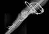
At 4 days after the emergency operation, we performed debridement again and inserted antibiotics (vancomycin)-mixed cement beads into the defect of the distal femur to prevent infection. The patient received 2nd-generation cephalosporin and aminoglycoside antibiotics during the initial 2 weeks and he received more 2nd-generation cephalosporin antibiotics during the following 2 weeks (Fig. 2).
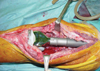
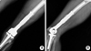
After 6 weeks, we performed a reconstructive total knee arthroplasty with modular segmental endoprosthesis, MUTARS® (Implantcast, Buxtehude, Germany) to treat the large bone loss of the distal femur (Fig. 3). Simultaneously, we carried out patellar tendon augmentation using a semitendinosus allograft because gracilis and semitendinosus were lost at the initial trauma (Fig. 4A). Furthermore, a medial gastrocnemius rotational flap with meshed skin graft was followed because conditions of the repaired patellar tendon and anterior skin of the knee were not healthy for rehabilitation of the knee joint (Fig. 4B).
One week after the reconstruction, physiotherapy team commenced continuous passive motion of the knee and crutch ambulation began 2 weeks after reconstruction.
At his most recent follow-up visit, 36 months postoperatively, the patient does not complain of pain, and can ambulate without support. The range of motion of the knee joint is 10 degrees to 55 degrees. Power of quadriceps muscle is 4/5, and the knee society knee score in pain is 79 and functional score is 50. The patient shows a mild limping gait because of 1.5 cm shortening of the left leg after the reconstruction arthroplasty. However, the patient can ambulate independently and is satisfied with the results (Fig. 5).
DISCUSSION
Popliteal artery is fixed to femur proximally by tendinous hiatus of adductor magnus and distally by a tendinous portion of soleus muscle. Therefore, it is susceptible to shearing or stretching by fractures of the lower femur and dislocations of the knee (1, 5, 6). Prompt recognition of associated popliteal artery disruption in damaged extremities and early revascularization are essential for good results as major factor favoring major limb salvage is a short preoperative ischemia time (2, 5, 6).
Massive trauma of lower extremity is particularly associated with vascular injuries and presents an immediate and complex decision-making challenge between limb salvage attempt and primary amputation. Lange (2) suggests two absolute indications that would warrant primary amputation in massive lower extremity trauma. The first is complete posterior tibial nerve disruption in adults; the second is crush injury with more than six hours of warm ischemic time. Unfortunately, the literature to date is unable to provide sound guidelines for treatment of such injuries. Individual patient variables, specific extremity injury characteristics, and associated injuries must all be weighed up before a treatment decision can be reached (2). Our patient had normal nerve function of the lower leg. In my opinion, this normal nerve condition would contribute to good long-term results.
In this case, multiple surgical options are available after revascularization, including arthrodesis, delayed amputation of a useless limb, allograft-prosthetic composite, and segmental prosthetic arthroplasty (2, 7, 8). Reconstruction methods to maintain a mobile knee joint are allograft-prosthetic composite, and segmental prosthetic arthroplasty (7, 8).
Modular segmental endoprosthesis with rotating-hinged knee kinematics was designed to allow reconstruction of large femoral deficits after tumor surgery (7). A modular segmental endoprosthesis is readily available and can easily be modified to fit the skeletal defect. This prosthetic arthroplasty also involves a shorter operation time, immediate weight-bearing with early stability, and early return to activities of daily living (4, 7).
Long-term survival results for endoprosthesis have not been well reported. There has been general reluctance to perform a total knee arthroplasty using constrained modular segmental endoprosthesis in young patients because of the variable results of a rotating-hinge knee design and potential complications, including aseptic loosening, infection, and component breakage (3, 4). Otherwise, Petrou et al. (9) reported good results in a primary total knee arthroplasty using Endo-model rotating-hinge prosthesis with a 96.1% survival rate at 15 yr postoperatively. Also, excellent long-term prosthetic survival for aseptic loosening was reported for 83 patients with a segmental rotating-hinge total knee arthroplasty of distal femur with 15-yr survival from the time zero point of 5 yr after the first operation noted to be 86% (7).
Another option, allograft prosthetic composite has many advantages, including biocompatibility, bone stock restoration, and potential for ligamentous reattachment. But disadvantages also exist; for example, late resorption of allograft possibly secondary to immune reaction, allograft fracture, nonunion, malunion, collapse, and the risk of disease transmission (7, 8). This procedure is also technically more difficult and would require a longer recovery and healing period for host-graft junction (4, 8).
We have described the case of severe knee joint injury asscociated with severe loss of the distal femoral bone and popliteal artery occlusion. Prompt recognition of artery disruption, early revascularization with intact function of posterior tibial nerve, and consecutive reconstruction surgery are important for successful and functional results (2, 7). Although we have used the modular segmental endoprosthesis, we would not advocate it as a routine procedure in cases when both allograft-prosthetic composite and modular segmental endoprosthesis were available. In our opinion, reconstructive surgery with modular segmental endoprosthesis was a suitable treatment option in terms of the short-term outcome in this case, where it was impossible to perform an internal fixation of fracture around the knee joint. However, we need to know and consider the long-term results of this case.

 XML Download