

 PDF
PDF ePub
ePub Citation
 Print
Print


INTRODUCTION
The idea that analgesic treatment initiated before the injury would be more effective than the same analgesic treatment given after the injury was named protective analgesia and showed various efficacies in many reports (1, 2). Transmission of pain signals evoked by tissue damage leads to sensitization of the peripheral and central pain pathways. Theoretically, protective analgesia using opioids or antihyperalgesic drugs such as N-methyl-D-aspartic acid (NMDA)-receptor antagonists and gabapentin, may interfere with the induction and maintenance of the sensitization. Therefore, immediate postoperative pain may be reduced, and the development of chronic pain may be prevented (1). Gabapentin (1-[aminomethyl]-cyclohexane acetic acid) has been shown to provide effective analgesia for various neuropathic pains. However, the use of gabapentin before the establishment of neuropathic pain has been rarely studied.
The α2δ1-subunit of the voltage dependent calcium channel (the α2δ1-subunit) is the only known binding site for gabapentin (3-5). The α2δ1-subunit is up-regulated in the dorsal root ganglion (DRG) and precedes the onset of neuropathic pain in a spinal nerve injury (6-8), and four days consecutive treatment of intraperitoneal gabapentin reduced the level of the α2δ1-subunit in the Chemotherapy-induced neuropathy (9).
Therefore, this study was designed to determine whether early gabapentin treatment before neuropathic pain establishment has a protective analgesic effect on neuropathic pain and compared its effect to the late treatment, and as the potential mechanism of protective action, the α2δ1-subunit was evaluated.
MATERIALS AND METHODS
These experiments were approved by the Institutional Animal Care and Committee of our Biomedical Research Institute, and were performed in accordance with the guidelines of the Committee for Research and Ethical Issues of IASP (10).
Animal preparation
Male Sprague-Dawley rats, weighing 130-180 g, were housed in separate cages with a 12/12 hr day/night cycle and food and water provided ad libitum. The rats were allowed to acclimatize for 5-7 days before the experiments.
Model of neuropathic pain
Neuropathic pain was induced using the procedure described by Kim and Chung (11, 12). Briefly, the rats were anesthetized with 2.5% enflurane in O2 through a mask. A dorsal midline incision was made from L3 to S2. Under microscopic guidance, the left L6 transverse process was partly resected to visualize the L4 and L5 spinal nerves. The left L5 spinal nerve immediately distal to the DRG was isolated and ligated tightly with a 6-0 black silk.
Gabapentin administration
Intraperitoneal gabapentin (Park-Davis, Ann Arbor, MI, U.S.A.) 30-300 mg/kg was reported to be an effective analgesic dose (7, 12-15). Gabapentin was dissolved in sterile saline 1 mL and injected intraperitoneally. One hundered mg/kg of Gabapentin was injected intraperitoneally 15 min before the nerve ligation (postoperative day [POD] 0) and then at 24 hr intervals during POD 1-4 in the early treatment group (n=21). For the late treatment group, the same dose of gabapentin was administered every 24 hr during POD 8-12 (n=21). The control group underwent the same nerve ligation but did not receive gabapentin (n=21).
Test for tactile allodynia
An observer who was blinded to the study design performed the tactile allodynia tests. Baseline values were obtained before nerve ligation. Test for tactile allodynia in all groups was carried at 8 a.m. everyday before next gabapentin administration to avoid diurnal variation.
For the baseline and POD tests, each rat was placed in a clear plastic cage (24×13×13 cm) with a 4×4-mm wire-mesh grid floor and allowed to acclimatize for at least 15 min. A series of von Frey hairs (number: 4.17, 4.31, 4.56, 4.74, 4.93, 5.07, and 5.18; Stoelting, Wood Dale, IL, U.S.A.) starting with 4.31 were applied in consecutive sequence through to the grid floor to the ventral surface of the operated hindpaw of each rat with a pressure causing the filament to buckle.
Brisk paw lifting within 5 sec indicated a positive response and prompted the use of the next weaker filament. An absence of a paw withdrawal response after 5 trials prompted the use of the next stronger filament. This paradigm was continued until five more measurements had been made after recording the initial change in the behavioral response.
50% gm threshold=10(Xf+κδ)/10,000
Where Xf=the value of the final von Frey filament used (in log units), κ=the tabular value for the pattern of positive/negative responses (from Chaplan et al. [17]), and δ=the mean difference between stimuli (in log units).
Rats showing any sign of motor dysfunction, including abnormal ambulation or placing/stepping reflex were excluded.
Harvesting the DRG
The DRGs were collected at POD 7, 14, and 21 in each group. Six rats in each group were euthanized in a CO2 gas chamber at POD 7, 14, and 21 after the surgery. The previous incision was reopened and widened. The L5 nerve was identified with a black silk tie and dissected carefully to the point of the DRG. The DRG was resected and preserved in a -70℃ refrigerator for Western blotting. The contralateral L5 DRG was also obtained for comparison. The level of the α2δ1-subunit in the DRG of nerve-ligated side was compared with the contralateral DRG and was expressed as the % of the contralateral side.
Western blot
The isolated DRG was homogenized with a glass Teflon homogenizer in an ice-cold homogenization buffer (10 mM Tris, pH 7.4, 1% Nonidet P-40, 150 mM NaCl, 0.1% SDS, andprotease inhibitors) and centrifuged at 20,000 g for 15 min. The supernatants were electrophoresed in 8% SDS-polyacrylamide gel and transferred to PVDF membranes (Millipore, Billerica, MA, U.S.A.). The membranes were incubated with a polyclonal antibody recognizing the α2δ1-subunit (cat#: AB5604-200 µL, Chemicon International, Temecula, CA, U.S.A.) or a monoclonal antibody recognizing the neuron specific beta III Tubulin (Abcam, Cambridge, U.K.) for 16 hr. After washing with Tris-buffered saline/Tween (0.05%), the blots were incubated with horseradish peroxidase-conjugated anti-rabbit, or anti-mouse IgGs, and developed using a LAS-3000 chemiluminescence imaging system (Fuji Film, Tokyo, Japan).
Data analysis
Kruskal-Wallis ANOVA on ranks and the subsequent post hoc comparisons (Dunn's multiple comparisons) were performed for allodynia and the level (%) of α2δ1-subunit between the three groups. A Friedman repeated measures ANOVA on Ranks (Tukey test for multiple comparison) was used to compare the time-dependent changes in allodynia. Chi-square test was used for % comparison of non allodynic rats between the groups. A p<0.05 was considered significant.
RESULTS
Allodynia
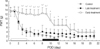
In the early treatment group, PWT did not decrease up to POD 10, whereas, in the control and the late treatment groups, PWT significantly decreased from the baseline value on POD 2 (p<0.05 compared to baseline value). PWT was higher in the early treatment group than in the control and the late treatment groups until POD 14 (p<0.05). The late treatment with gabapentin failed to show any significant anti-allodynic effect (Fig. 1).
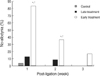
The 83.3% of rats in the early treatment group did not develop allodynia on POD 7, which was significantly higher than in the control (3.3%) and the late treatment group (12.5%) (p<0.05). On POD 14, the 36.4% of rats in the early treatment group did not develop allodynia, which was still higher than in the control (0%) and the late treatment groups (8.3%) (p<0.05). On POD 21, 16.7% in the early treatment group did not develop allodynia, whereas all the rats developed allodynia in the control and the late treatment group (p<0.05) (Fig. 2).
Expression of α2δ1-subunit
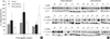
The α2δ1-subunit in the ligated side DRG was up-regulated in all groups (p<0.05).
However, the level of the α2δ1-subunit between the three groups at 1, 2, and 3 weeks after nerve ligation were not different (Fig. 3).
DISCUSSION
In the present study, the early gabapentin treatment produced a significant anti-allodynic effect, whereas this effect was absent in the late gabapenitn treatment. However, gabapentin treatment did not affect the level of the α2δ1-subunit in DRG.
Protective effect for neuropathic pain by gabapentin has been shown in a few recent studies. Gabapentin when given before hysterectomy reduced postoperative pain (18). Kaneko et al. (19) showed that pre-treatment of gabapentin was three times more potent in pain relief than post-treatment in formalin hindpaw injection. This was a postoperative pain or formalin injection study with single intrathecal gabapentin administration. For neuropathic pain, Coderre et al. (20) reported that the early consecutive treatment with gabapentin, but not the late treatment, showed anti-allodynic effect in a chronic constriction injury of sciatic nerve. In their study, rats were injected with gabapentin (30, 100 or 300 mg kg-1) intraperitoneally 15 min prior to the surgery and then during POD 1-4 (pre-treatment group), or during POD 8-12 (post-treatment group) at 12 hr intervals. They showed that the early treatment has an anti-allodynic effect compared to the vehicle injection, whereas this effect was not evident in the late treatment group.
In our study, the anti-allodynic effect of gabapentin in the early treatment group persisted even after discontinuing the drug. In contrast, the late treatment with gabapentin did not have any apparent efficacy against allodynia. Peak analgesic effect of gabapentin appears at 1-2 hr after intraperitoneal injection and decreases to pre-injection level within 4-5 hr (9, 12, 15, 21). Therefore, the persistent anti-allodynic effect in the early treatment group, even after discontinuing the drug, might imply some type of structural change in the nervous system.
In our study, the level of the α2δ1-subunit in DRG was not different among the groups. There was only one study that dealt the effect of gabapentin treatment on the level of the α2δ1-subunit (9). They administered gabapentin during the peak neuropathic pain period in a chemotherapy induced neuropathy model and compared 1 day-gabapentin treatment with 4 days-treatment (n=7, each group) revealing that only repeated dosing was effective in reducing allodynia and the α2δ1-subunit in dorsal spinal cord. Interestingly, in their study, no up-regulation of the α2δ1-subunit or reduction by gabapentin treatment, was shown in the DRG. Our study dealt with nerve ligation induced neuropathy and measured the α2δ1-subunit in the DRG. In the case of nerve ligation, up-regulation of the α2δ1-subunit was much higher in the DRG than in the dorsal spinal cord (6, 8). In addition, we compared the early and late treatment with gabapentin, not the duration of treatment. These differences of the study design might have resulted in different outcomes, however, the α2δ1-subunit in the dorsal horn need to be evaluated in a further study.
Excitatory amino acids such as NMDA (20, 22) and their receptors (23), gamma-aminobutyric acid receptors (24, 25), norepinephrine (26, 27), and K+ channels (27, 28) have been suggested to involve in analgesic mechanisms of gabapentin. However, these mechanisms have not been verified for the protective effect of gabapentin yet. Among them, protective effect of NMDA antagonist is well known (29, 30), and NMDA receptor antagonist attenuated the up-regulation of the NMDA receptor subunit when administered before a nerve injury (29). The α2δ1-subunit enhances the Ca2+ channel currents (31, 32), and longer depolarization and concomitant calcium entry facilitate the release of glutamate and substance P from the nerve ending. This activates the NMDA receptors and results in a wind-up phenomenon (33). Gabapentin by blocking the α2δ1-subunit might prevent the whole cascade of subsequent events. Future studies are needed to examine the mechanisms of the protective role of gabapentin.
In the early treatment group, allodynia finally developed after 2 weeks. The only way of preventing the sensitization of the nociceptive system might be to block all pain signals originating from the surgical wound completely from the time of the incision until the final wound healing (1). Xie et al. (34) also reported that a shorter duration of the blockade of nociceptive input is one of the reasons for the pre-treatment failure. Therefore, further study is needed to find that a longer treatment of gabapentin that can cover the whole period of the noxious peripheral input could prevent the development of allodynia.
We started gabapentin before nerve injury for protective analgesia, however, it is hardly the case in clinical situation except very high probability of development of neuropathic pain such as limb amputation. Therefore, gabapentin treatment shortly after nerve injury and before neuropathic pain development might give valuable information on the timing of treatment.
In conclusion, the results of our study demonstrated a protective analgesic effect of gabapentin in a L5 ligation model. Administration of gabapentin before the neuropathic pain establishment showed long lasting anti-allodynic effect. However, the mechanism is not related with the suppression of the up-regulation of the α2δ1-subunit in DRG. Our result implies that early administration of gabapentin after injury to the patients who have a high probability to develop neuropathic pain would be beneficial. Humane studies and further steps to elucidate underlying mechanisms are essential for gabapentin-induced protective analgesia.
 XML Download