

 PDF
PDF ePub
ePub Citation
 Print
Print


INTRODUCTION
Congenital middle ear cholesteatoma (CMEC) manifests as a whitish mass in the middle ear with intact tympanic membrane in young ages, and it is a relatively rare disease consisting of approximately 2% of the entire cholesteatoma cases (1). Recently, with the increase of interests on this disease, its detection rate is increased, and thus the ratio of congenital cholesteatoma to the entire cholesteatomas has been reported to be from 3.7% to 24% (2).
The symptoms are diverse depending on the location and the extent of lesions from asymptomatic to the conductive hearing loss, labyrinthitis, facial palsy, sensorineural hearing loss, intracranial complications, etc. The representative hypotheses of the etiology are the defect of the tympanic ring, epidermoid formation, epithelial metaplasia and implantation theory (2-4). Nevertheless, none of the hypotheses explain accurate mechanisms. Recently, together with studies on the progression of congenital cholesteatoma, their classification has been reported. Koltai et al. (5), Nelson et al. (6), and Potsic et al. (7) have suggested the disease stage based on the presence or absence of the invasion to the ossicle and the invasion to the mastoid.
In the treatment of CMEC, it is very important to classify the pattern of cholesteatoma and to determine the lesion according to it, and to evaluate accurately the damage of important anatomical structures and complications prior to surgery. It is thought that the prognosis may be different depending on the location of CMEC, its disease stage, and surgical methods. Therefore, in pediatric CMEC underwent surgical treatment, we analyzed the association of the computed tomography (CT) stage, the disease stage at the time of surgery (surgical stage), surgical methods, the result of audiologic test and recurrence rate.
MATERIALS AND METHODS
On 35 pediatric CMEC who were treated surgically in the department of otolaryngology head and neck surgery at the Catholic university hospital from 1995 to 2006. Follow up period were ranged from 9 to 128 months, average 47 months. And we reviewed clinical symptoms, physical findings, audiologic test, radiologic test and surgical findings retrospectively.
The patients were children younger than the age of 15 yr, and the diagnosis of CMEC was made based on the diagnostic criteria suggested by Levenson et al. (8) that 1) a whitish mass present in the middle ear cavity with normal tympanic membrane, 2) the pars flaccida and pars tensa of tympanic membrane showed normal findings, 3) without the past history of otorrhea and perforation, 4) without the history of otological surgery, 5) the occulusion of the external canal, intramembranous cholesteatoma and giant cholesteatoma were excluded, and 6) the past history of otitis media was not precluded.
Regarding the classification of CMEC according to their location, based on the handle of malleus, it was divided into the lesion in the anterior and posterior area.
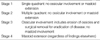
As the disease stage of cholesteatoma, based on the classification suggested by Potsic et al. (7), the CT stages and surgical stages were compared (Tabel 1). Depending on the disease stage, tympanoplasty, epitympanoplasty, canal wall up mastoidectomy, canal wall down mastoidectomy, and if needed, ossiculoplasty was performed.
As audiologic test, according to the guideline of the American Academy of Otolaryngology Head and Neck Surgery (1995), the mean hearing level at 500 Hz, 1,000 Hz, 2,000 Hz, and 3,000 Hz was used. The analysis of hearing test after surgery was performed based on the most recent audiologic test, 2-3 months after the surgery. The hearing change after surgery was defined as the difference of the air bone conduction threshold gap prior to surgery and the air bone conduction threshold gap after surgery.
The statistical significance of the hearing change after surgery was assessed by paired t-test and Wilcoxon signed rank test, and a significant level was p<0.05.
RESULTS
The study population
The age of patients was from 2 to 13 yr old and their mean age was 6.2. The boy was 27 patients, the girl was 8, and the ratio of the male and the female was 3.4:1. CT stage according to Potsic et al. (7), the stage 1 was 2 patients, the stage 2 was 4, the stage 3 was 10, and the stage 4 was 17. The mean age at the time of surgery in the stage 1 was 4.5 yr old, the stage 2 was 6.3, the stage 3 was 5.1, and the stage 4 was 7.3.
Clinical symptoms and physical findings
All patients had a unilateral lesion, developed in the left side was in 15 cases (43.0%), and in the right side in 20 (57.0%). Asymptomatic cases discovered were 16 cases (45.0%). And in symptomatic cases, hearing impairment was 10 (29.0%), which was most prevalent, otalgia was 7 (20.0%), tinnitus was 4 (11.4%), and ear fullness was 3 (8.5%), and in others 1 case (2.8%), a symptom similar to postauricular swelling. In the past history, the cases who had acute otitis media or otitis media with effusion were 11 patients (31.4%). Concerning the otoscopic findings, normal findings were 7 patients (20.0%), a whitish mass in the normal eardrum was detected in 21 (60.0%), and bulging of posterior part of tympanic membrane was detected in 7 (20.0%), and none of patients had a perforation in the tympanic membrane.
Audiologic test
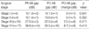
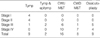
Among 35 patients, preoperative audiologic test was performed on 23 patients. The air bone conduction threshold gap prior to surgery in the CT stage 1 & 2 was 10.0 dB, in the stage 3 was 27.0 dB, in the stage 4 was 37.5 dB, and a trend of the increase with advanced disease stage was detected. Patients underwent audiologic test prior to surgery and 2-3 months after surgery in the stage 3 were 4 cases and the stage 4 was 10. The improvement of the air bone conduction threshold gap in the stage 3 was 2.5 dB and the stage 4 was 6.7 dB, but statistically significant improvement was not detected (Table 2).
Radiological test
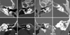
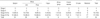
Based on the disease stage by Potsic et al. (7), 35 patients were classified according to their disease stage based on preoperative CT findings (Fig. 1) and surgical findings, and compared. It was found that the cases whose CT stage corresponding to surgical stages were 30 cases (85.7%), the cases whose CT stage was underestimated in comparison with surgical stages were 2 (5.7%), and the overestimated cases were 3 (8.6%) (Table 3).
Surgical findings & surgical methods
Regarding the disease stage at the time of surgery, the surgical stage 1 was 4 cases (11.4%), the stage 2 was 4 (11.4%), the stage 3 was 10 (28.6%) and the stage 4 was 17 (48.6%).
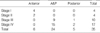
4 cases of the surgical stage 1 were restricted to the anterosupeior area, and 2 cases of the stage 2 were limited to the anterior area and the others of the stage 2 were limited to posterior area. It was found that in 26 cases of the stage 3 and 4, none of cases was limited to the anterior area, the posterior area was 3 cases, and the both anterior and postrior was 24 (Table 4).
In the stage 3 and 4 invaded to more than the middle ear cavity, the cases invaded up to the epitympanum was 9 cases (90.0%) in the stage 3 and 15 cases (88.2%) in the stage 4, and the cases expanded to the hypotympanum in the stage 3 was 4 cases (40.0%) and 8 cases (47.0%) in the stage 4. The invasion to the sinus tympani in the stage 3 was 1 case (10.0%) and 7 cases (41.1%) in the stage 4. Invasion to the facial recess in the stage 3 was 3 cases (30.0%) and 7 cases (41.1%) in the stage 4. And invasion to the Eustachian tube in the stage 3 was 4 cases (40.0%) and the stage 4 was 7 cases (41.1%). As the stage was advanced, the invasion to the sinus tympani, the facial recess and the Eustachian tube was increased (Table 5). In the cases of invasion into the facial recess and sinus tympasni, we tried to remove cholesteatoma completely using many microinstruments, especially with micromirror.
Regarding the invasion into the ossicles, the malleus was 9 cases, the incus was 26 and the stapes was 20. The damage of the facial canal in the stage 3 was 1 case and the stage 4 was 6, the dura exposure in the stage 3 was 1 and the stage 4 was 2, and none of patients had the exposure in the lateral semicircular canal. As the disease stage is advanced, the exposure of facial canal or dural exposure becomes more frequent (Table 6).
Regarding the surgical procedures, tympanoplasty was done for the patients with stage 1 & 2. And there was no recurrence. As for the stage 3, tympanoplasty was done for 1 case, tympanoplasty & epitympanoplasty for 2 cases, canal wall up mastoidectomy with tympanoplasty for 6 cases and canal wall down mastoidectomy with tympanoplasty for 1 case. And ossiculoplasty was done at the time surgery for 5 cases (5/10) with partial ossicular replacement prothesis (PORP). As for stage 4, canal wall up mastoidectomy with tympanoplasty was done for 10 cases and canal wall down mastoidectomy with tympanoplasty for 7 cases. Ossiculoplasty was done at the time of the first surgery for 4 cases (4/17) using PORP.
Recurrence
It recurred in 2 cases among 35 patients (5.7%). They were all the surgical stage 4 and underwent canal wall up mastoidectomy. As revisional surgery, canal wall down mastoidectomy was performed in 1 case, and revision canal wall up mastoidectomy was performed in the other. Ossiculoplasty was performed in all cases. At the time of revisional surgery, in all 2 cases, the invasion of cholesteatoma in the sinus tympani and the facial recess were observed, and they were the patients who had the invasion to the sinus tympani and the facial recess also at the first surgery. Until the last follow up observation, findings suspicious of recurrence were not detected in 33 patients, and the second look operation was not performed.
DISCUSSION
House (9) reported the case of CMEC for the first time in 1953, and in 1965, Derlacki and Clemis (10) established the diagnostic criteria for congenital cholesteatoma. Levenson et al. (8) confirmed the diasease history of acute otitis media in 70% of infants younger than the age of 2 yr in the U.S.A. and thus did not preclude the past history of acute otitis media from the diagnostic criteria. Similarly, we did not preclude the past history of otitis media according to the diagnostic criteria of Levenson et al. (8) and patients who had acute otitis media or otitis media with effusion were 11 cases (31.4%), which was not greatly different from the previous report 9-55% (11).
Clinical symptoms of CMEC manifest diversely depending on the advanced stage. Generally, asymptomatic cases are most prevalent and the symptoms such as conductive hearing loss, ear fullness, otalgia, tinnitus, dizziness, headache, paralysis of facial nerves, etc. can be detected. House and Sheehy (12) have reported that in the cases with the unilateral conductive hearing loss which is difficult to explain by unilateral otitis media with effusion, regardless of the age of patient, the possibility of CMEC should be considered. In our study, asymptomatic cases discovered incidentally were 45%, and the cases who did not show any specific findings with otoscope were 20%. And at the time of surgery, the advanced lesion equivalent to the disease stage 3 or 4 was detected in most cases. In addition, it was found that as the disease stage become advanced, the frequency of the invasion to the epitympanum, the hypotympanum, the sinus tympani, the facial recess, the facial canal and the dura was increased. Considering that CMEC is discovered frequently in its advanced stage, it is thought that for its early diagnosis, a screening program including otoscopic examination, hearing test and CT for children may be required.
As the etiology of CMEC, defect in the tympanic ring, epidermoid formation, epithelial metaplasia and implantation theory have been proposed. Nonetheless, none of hypotheses could explain the etiology completely (2-4).
Concerning the location of CMEC, Levenson et al. (8) have reported that it is located primarily in the anterior area of the tympanic cavity and Karmarkar et al. (13) have reported that the posterior area of the tympanic cavity is the site where it is developed preferentially. But recently, it has been reported to be developed in the posterior area of the tympanic cavity more frequently (11). In our study, among 35 cases, 6 cases (17.1%) were limited to the anterior area, 5 cases (14.2%) were limited to the posterior area, and in 24 cases (68.5%), the anterior and posterior area were involved simultaneously.
Nelson et al. (6) classified CMEC according to the presence or absence of the invasion to the ossicles and the mastoid. The lesion limited to the middle ear is the type 1, with the invasion to the ossicles is the type 2, invasion to the mastoid is the type 3. And toward to the type 3, its recurrence was increased. Potsic et al. (7) and Kazahaya and Potsic (14) have reported that in the classification according to Nelson et al. (6), the cases without invasion to the ossicle were divided again into two groups and classified to total 4 groups.
The risk of residual cholesteatoma after surgery in the disease stage 1 was 13% and the stage 4 was 67%. Hence, the risk of residual cholesteatoma was increased as the disease stage advanced, and proposed the disease stage system that could predict the prevalence rate of residual cholesteatoma.
Surgical methods vary depending on the progression of the lesion. According to Nelson et al. (6), the type 1 could be treated sufficiently only by tympanoplasty, the type 2 may require extended tympanoplasty, canal wall up mastoidectomy, ossiculoplasty, and second look operation is required occasionally. And the type 3 are similar to type 2 cases, nonetheless, it may require canal wall down mastoidectomy. However, canal wall down mastoidectomy has a shortcoming that continuous ear treatment is required throughout the life and the possibility of developing cavity problem is high. And thus it should be performed only in the cases with the defect in the posterior wall of the external auditory canal, invasion of cholesteatoma into the inner ear or the petrous apex, and the cases whose follow ups are difficult (15). Ueda et al. (16) also recommended to perform canal wall up mastoidectomy if possible, and to choose the one-stage surgery for the cases that on presurgical CT findings, the epitympanum and the mastoid were aerated, and without the damage in the malleus and the incus, to perform sequential tympanoplasty. Because such principle increases the possibility of the aeration of the epitympanum and the mastoid, if residual cholesteatoma could be detected early by CT, the formation of an attic retraction pocket could be prevented. Gocmen et al. (17) also recommended the canal wall up mastoidectomy for limited lesions and the canal wall down mastoidectomy for cholesteatoma in a wider area.
The results of recurrence rate, improvement of hearing and hearing deterioration were not different between the cases performed the wall down mastoidectomy or the wall up mastoidectomy. In our cases of advanced stage, we did one stage operation with ossiculoplasty. We thought that we removed the cholesteatoma properly and tried to reduce hearing difficulty of patients and concerns of second operation.
The rate of residual cholesteatoma at the time of second surgery is very diverse, 8-81% (18). In our study, recurrence was observed 2 cases (5.7%) and a relatively low recurrence rate was shown, which is thought to be the reason that the number of patients was 35 cases which was not large, and the follow-up period in some patients was not long. Two recurred cases were the disease stage 4 at the time of first surgery, with the invasion to the sinus tympanic and the facial recess, and the canal wall up mastoidectomy was performed. And at the time of recurrence, the recurrence in the sinus tympani and facial recess could be observed. It is considered that such result was obtained due to the failure of the complete removal of cholesteatoma by the canal wall up mastoidectomy. And it is thought that for the cases with invasion in these areas, to prevent recurrence, the canal wall down mastoidectomy is required.
To perform CT prior to surgery considered as a standard preoperative examination nowadays. Levenson et al. (8) accepted the usefulness of CT as presurgical test, nevertheless, they preferred micro-otoscopy. Friedberg (2) have reported that imaging tests were a great help to understand the extent of lesion and to decide surgical approach. And El-Bitar et al. (19) claimed that CT is of help to understand the extent of cholesteatoma and to evaluate invasion to the ossicles, and mediates an effect on surgery outcomes. By using CT, Kim et al. (20) detected the invasion to the middle ear cavity in 80%, invasion to the mastoid in 76.5%, and invasion to the sinus tympani in 50%. And as the prediction of the ossicle destruction, the malleus, the incus and the stapes was shown to be 75%, 65%, and 85%, respectively. In our study, the result showed that the CT stage corresponded to surgical stage in 85.7%. It is thought that by predicting the location of lesion and the spread route by CT imagings prior to surgery, it is of help to plan the treatment methods and reduction of complications for cholesteatoma patients.
In pediatric congenital middle ear cholesteatoma, it is thought that screening tests including CT for its early detection are required. Considering the preoperational evaluation, CT showed a high corresponding rate to the surgical stage determined by Potsic. CT is thought to be a useful tool for the presurgical evaluation of cholesteatoma, its treatment method and follow ups. In the cases that the cholesteatoma was large and invaded to the sinus tympani or the facial recess, the canal wall down mastoidectomy may be required to reduce recurrence.


 XML Download