

 PDF
PDF ePub
ePub Citation
 Print
Print


INTRODUCTION
Lower urinary tract symptoms (LUTS) secondary to benign prostatic hyperplasia (BPH) are one of the most common disorders in elderly males, and alpha1-blockers are now viewed as first-line agents for the management of LUTS caused by BPH. Alpha1-blockers originally were believed to work by blocking alpha adrenergic receptors found in the prostate and its capsule, and thereby relaxing stromal smooth muscle (1). However, this mechanism does not fully account for the long-term clinical responses observed for these drugs in BPH. Moreover, recent studies have demonstrated that alpha1-blockers, such as doxazosin and terazosin, induce apoptosis of prostate stromal smooth muscle and epithelial cells without affecting cellular proliferation, and have correlated this with symptom improvement (2-4). It was also found that the apoptotic effects of doxazosin and terazosin are mediated by a mechanism involving the quinazoline nucleus and not one involving alpha1-adrenoceptor blockade, since the non-quinazoline alpha1-adrenoceptor antagonist tamsulosin does not elicit an apoptotic response (5).
The apoptosis of prostate tissue induced by alpha1-blockers may confer a considerable benefit in BPH, since it could alter the size and shape of the prostate over time, reduce its volume, or halt further growth. Moreover, previous studies have shown that 5alpha reductase inhibitor reduces prostate size and serum prostate-specific antigen (PSA) levels by inducing apoptosis (6, 7). However, few systematic studies have been undertaken to test this hypothesis, although some investigators have reported that alpha1-blockers do not affect serum PSA concentrations (7, 8). We conducted the present study to compare the impacts of terazosin and tamsulosin on prostate activity. In this study, the serum PSA or prostate volume were taken to reflect prostate activity. In fact, PSA was used recently as an indicator of prostate gland activity and function (9-11).
MATERIALS AND METHODS
The clinical records of patients who had presented with LUTS secondary to BPH between January 2003 and December 2005 were retrospectively reviewed. The study inclusion criteria were: age ≥50 yr, moderate to severe LUTS (International Prostate Symptom Scores [I-PSS] sum ≥8), and the ability to communicate, understand, and comply with the requirements of the study. The exclusion criteria included: the use of other medications to control bladder symptoms, the presence of a bladder tumor, bladder stone, or urethral stricture, neurogenic bladder dysfunction, and restricted mobility. Patients were also excluded from the analysis if they had prostate cancer or prostatic intraepithelial neoplasia by biopsy, a serum PSA level of >20 ng/mL, a history of prostate surgery or radiotherapy, an acute urinary retention or an indwelling catheter, evidence of an acute urinary infection (pyuria and bacteriuria) on urine analysis, or a medication history of alpha1-blocker or 5alpha-reductase inhibitor. Ninety patients met the criteria and constituted the study cohort. The median (range) age of the study patients was 65.0 (52.0-83.0) yr, their median baseline total prostate volume (TPV) and transition zone volume (TZV) were 31.5 (17.0-88.0) and 9.6 (2.0- 45.2) mL, respectively, and their median baseline PSA level was 1.2 (0.2-14.8) ng/mL.
At initial visits, patients underwent a detailed clinical evaluation, including a complete history taking, a physical examination, urinalysis, urine culture, and a digital rectal examination (DRE). In addition, the followings were measured; PSA level, prostate volume by transrectal ultrasonography (TRUS), uroflowmetry, and post-void residual (PVR) urine volume. I-PSS were also determined. To determine TPV and TZV, TRUS was performed by a single radiologist using a 7.0 MHz. transducer (Ultramake-9, ALT Inc., Washington DC, U.S.A.). Subjects were placed in the left lateral decubitus position with knees pulled up toward the chest. After DRE, the probe was inserted and Gray scale ultrasonography was performed. Anteroposterior (H) and transverse prostate diameters (W) were measured on largest transverse images. Horizontal distances between the most proximal and distal prostate boundaries on midline sagittal scans were considered to be longitudinal diameters (L). Prostate volumes were estimated assuming an ellipsoid shape using the formula, prostate volume=π/6×H×W×L (12). Patients aged ≥50 yr with suspicious results on DRE and/or a high PSA level (>4 ng/mL) also underwent a systemic sextant biopsy, under TRUS guidance, using an 18-G needle fitted to an automatic biopsy gun. Hypoechoic lesions detected by ultrasonography and areas corresponding to palpable abnormalities on DRE were also biopsied.
Patients were given 0.2 mg tamsulosin, or 2 mg or 4 mg terazosin once daily. The median treatment duration was 14.0 (6.0-56.0) months. Uroflowmetry was conducted, with measurements of PVR via abdominal ultrasonography. Maximum flow rates (Qmax), voided volumes, and PVR were assessed pre- and post-treatment. Bladder voiding efficiency (BVE) was defined as voided volume (determined by uroflowmetry) divided by initial urine volume (voided volume plus PVR) (13);
BVE=(voided volume/total bladder capacity)×100
Prostate volume was measured by TRUS at the end of the treatment period. LUTS and symptom-specific quality of life (QOL) were assessed using I-PSS and associated QOL index score pre- and post-treatment.
Survey responses were coded and analyzed using descriptive statistics, and were reported as medians (25-75th percentiles) or as numbers and percentages (qualitative variables). For statistical analysis, patients were categorized according to the medication administered, i.e., '0.2 mg tamsulosin' (n=19) and '2 mg terazosin' (n=16) and '4 mg terazosin' (n=55) or according to serum PSA, TPV or TZV reductions. Statistical analysis was carried out using the Mann-Whitney U test or the Kruskal-Wallis test for continuous data and the Armitage test for categorical data. When a significant group difference was found using the Kruskal-Wallis test, Turkey's multiple comparison test was performed to determine significance.
To identify factors associated with serum PSA, TPV, or TZV reduction, we calculated odds ratios (ORs) and p values for trends using univariate and multivariate logistic regression analyses. Clinical characteristics (age, serum PSA levels, TPV, TZV, type of alpha1-blockers, and treatment duration) were individually entered into a univariate model, and variables with a significance threshold set at p<0.100 were entered into a multivariate model as dependent variables. In our logistic regression model, the variables, age, serum PSA, TPV, TZV, and treatment duration were dichotomized about median values. The associations between clinical variables and serum PSA, TPV, and TZV reductions were determined using maximum likelihood estimates of relative risk with 95% confidence intervals (CIs). CIs were predicated from standard errors of measurement (SEMs) of coefficients, assuming a normal distribution.
A 5% level of significance was used throughout, except as mentioned above for the univariate logistic regression analysis, and all statistical tests were two-sided. Statistical analyses were performed using a commercially available program, SPSS 10.0 (SPSS, Inc., Chicago, IL, U.S.A.).
RESULTS
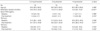
The baseline and post-treatment group prostate activities are shown in Table 1. At baseline, no differences were evident between the three groups with respect to age, serum PSA, TPV or TZV, although treatment durations were significantly different (p<0.001). After treatment, groups serum PSA and TZV values were similar, but TPV was smaller in the 2 mg terazosin group than that in other two groups (p=0.017). After treatment, 10.5% (2 of 19) in the 0.2 mg tamsulosin group showed a TZV reduction, whereas 37.5% (6 of 16) in the 2 mg terazosin group and 43.6% in the 4 mg terazosin group showed a TZV reduction (p=0.010).
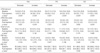
Table 2 provides details of outcomes versus changes in prostate activity, and shows that I-PSS total scores and QOL index score decreased regardless of prostate activity after treatment. However, objective parameters, namely, Qmax, PVR, and BVE, were not different after treatment, and were at similar levels in the three groups.
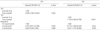
Logistic regression analysis was used to identify predictors of a prostate activity reduction (univariate analysis failed to identify any predictor of PSA reduction), and a reduction in TPV was found to be associated with baseline TPV and the medication administered. To exclude the possibility of a confounding effect, these two variables were subjected to multivariate logistic analysis, and baseline TPV was found to be the only independent predictor of a TPV reduction. Specifically, a baseline TPV of ≥30 g was found to have an OR of 3.939 for a TPV reduction (95% CI, 1.506-10.304; p=0.005).
On the other hand, univariate analysis indicated the likelihood of a TZV reduction was dependent on two factors, i.e., baseline TZV and the medication administered, while multivariate analysis showed that a baseline TZV of ≥10 g was associated with a 7.1-times greater chance of a TZV reduction (OR, 7.100; 95% CI, 2.428-20.763; p<0.001). Using the same model, patients on 2 mg terazosin were found to have a 10.8-fold greater chance of a TZV reduction (OR, 10.770; 95% CI, 1.409-82.323; p=0.022), and patients on 4 mg terazosin a 9.0-fold greater chance (OR, 9.001; 95% CI, 1.724-46.995; p=0.009) of a TZV reduction than those on 0.2 mg tamsulosin. These results are summarized in Table 3.
DISCUSSION
The prostate is richly innervated with autonomic nerves, which include both adrenergic and cholinergic fibers. The prostate expresses four native alpha1-adrenoreceptors, and one of these, alpha1a, is known to be related to smooth muscle contraction (14). Pharmacologic blockade of alpha1-adrenergic receptors relaxes prostate and bladder neck smooth muscle and improves the symptoms of BPH, and available data support the notion that quinazoline alpha1-adrenoceptor antagonists, such as doxazosin and terazosin, inhibit prostate growth via smooth muscle cell apoptosis. Moreover, doxazosin and terazosin are known to induce prostate smooth muscle cell apoptosis via mechanisms unrelated to alpha1-adrenoceptor blockade (15), and since smooth muscle represents approximately 40% of BPH tissue (16), smooth muscle apoptosis and the resulting stromal regression provide a cellular basis for the therapeutic effects of an alpha1 blockade.
Transforming growth factor (TGF)-beta1 has potent antiproliferative and cytotoxic effects on human benign prostatic epithelial cells (17). Moreover, recent experimental evidence points to the deregulation of signal transduction pathways involving TGF-beta and the disruption of cell attachment to the extracellular matrix as potential mechanisms that underlies the apoptotic effect of quinazoline-based alpha1-adrenoceptor antagonists on prostate cells (4).
In addition, experimental and clinical evidence indicates that the induction of prostate smooth muscle cell apoptosis by doxazosin or terazosin contributes to the long-term improvements observed in LUTS in patients with BPH. In fact, one study found a direct correlation with borderline significance (r=0.18, p<0.05) between doxazosin-mediated prostatic smooth muscle cell apoptosis and the improvement of BPH symptoms (3). Moreover, this is consistent with the findings of another study, which demonstrated a direct relationship between clinical response to alpha1-adrenoceptor antagonist therapy and smooth muscle cell proportion in BPH tissue (18).
Recently, Roehrborn (19) examined whether 3 months of alfuzosin treatment changes either TPV or TZV, and found that both were unaffected after 3 months of treatment. Although apoptotic induction by alfuzosin was not tested in a clinical study, its quinazoline structure suggests that it may induce similar apoptotic effects. However, since 3 months' treatment with alfuzosin may be a short treatment period to induce volume changes, additional studies are required to determine the effect of alpha1-blockers on prostate apoptosis and volume in patients over longer treatment periods (19).
In the present study, we failed to find any significant treatment-related differences in serum PSA, TPV, and TZV for 0.2 mg tamsulosin, 2 mg terazosin, or 4 mg terazosin. Nevertheless, larger baseline TPV and TZV values were found to be associated with an increased likelihood of a treatment-related TPV or TZV reduction, respectively. Furthermore, terazosin was found to be more likely to reduce TZV than tamsulosin. However, symptom improvement was not found to be associated with reduced prostate activity. Our findings suggest that the quinazoline alpha1-blocker terazosin influences prostate volume (especially TZV), and demonstrate that relief from the symptoms of BPH may not be due to prostate volume reductions induced by alpha1-blockers within the gland.
In the present study, no prostate tissue biopsy specimens were obtained, and it is evident that only direct identification of apoptotic cells in appropriate prostate tissue biopsy samples is likely to show definitively whether apoptotic induction is directly associated with serum PSA, prostate volume reductions, and symptom relief. Although it has been reported that tamsulosin does not induce prostate cell apoptosis in vitro (15), prostate tissue from tamsulosin-treated patients has not been examined for the presence of apoptotic cells after several months of treatment, which leaves the possibility open that in vitro and in vivo differences exist (19). Moreover, if tamsulosin does not elicit an apoptotic response, smooth muscle relaxation alone does not fully account for its long-term clinical effect. Thus, a study on a larger population for a longer period of observation is required to determine the clinical significances of terazosin and tamsulosin-induced apoptosis and to characterize their alpha1-dependent and -independent contributions.
 XML Download