Journal List > J Korean Med Sci > v.23(3) > 1020664

Choi, Kwon, Kim, Quinn, Hales, and Seo: Atelectasis Induced by Thoracotomy Causes Lung Injury during Mechanical Ventilation in Endotoxemic Rats

Abstract

Atelectasis can impair arterial oxygenation and decrease lung compliance. However, the effects of atelectasis on endotoxemic lungs during ventilation have not been well studied. We hypothesized that ventilation at low volumes below functional residual capacity (FRC) would accentuate lung injury in lipopolysaccharide (LPS)-pretreated rats. LPS-pretreated rats were ventilated with room air at 85 breaths/min for 2 hr at a tidal volume of 10 mL/kg with or without thoracotomy. Positive end-expiratory pressure (PEEP) was applied to restore FRC in the thoracotomy group. While LPS or thoracotomy alone did not cause significant injury, the combination of endotoxemia and thoracotomy caused significant hypoxemia and hypercapnia. The injury was observed along with a marked accumulation of inflammatory cells in the interstitium of the lungs, predominantly comprising neutrophils and mononuclear cells. Immunohistochemistry showed increased inducible nitric oxide synthase (iNOS) expression in mononuclear cells accumulated in the interstitium in the injury group. Pretreatment with PEEP or an iNOS inhibitor (1400 W) attenuated hypoxemia, hypercapnia, and the accumulation of inflammatory cells in the lung. In conclusion, the data suggest that atelectasis induced by thoracotomy causes lung injury during mechanical ventilation in endotoxemic rats through iNOS expression.

INTRODUCTION

Atelectasis is frequently encountered in clinical practice during general anesthesia, the post-operative period (1, 2), and when suctioning during mechanical ventilation (3). Atelectasis has been reported to occur in 23% of patients after surgical treatment of esophageal cancer (4). The incidence of acute lung injury (ALI) has been reported to be as high as 23.8% in certain types of elective surgery (5). Taken together, atelectasis may be one of the contributing factors of lung injury seen in these patients.
Many studies suggest that atelectasis induced by surfactant depletion or inactivation is injurious (6-8). However, atelectasis induced by the reduction of lung volume may have a different effect on lung injury. Atelectasis induced by thoracotomy and lung over-inflation increased pulmonary vascular permeability and induced inflammatory gene expression both in an in-vivo and an ex-vivo model (9, 10). However, the effect of atelectasis on ventilation with conventional tidal volumes has not been explored, and this is important since large tidal volume ventilation alone induces inflammatory cell infiltration (11).
Although surfactant dysfunction with alveolar collapse may be an important pathogenic mechanism in ALI, we suggest that lung volume loss itself may accelerate ALI during sepsis. Therefore, we hypothesized that lung volume below the normal functional residual capacity (FRC) would predispose the lungs to develop inflammation.
Shear stress stimulates inducible nitric oxide synthase (iNOS) expression in cultured smooth muscle cells (12). Endotoxemia and large tidal volume ventilation also induce iNOS expression, neutrophil infiltration, and increased microvascular permeability (13-15). Taken together, endotoxemia in combination with shear stress induced by low lung volume ventilation would have the potential to promote significant iNOS expression.
To test our hypothesis, rats were pretreated with lipopolysaccharide (LPS) to mimic sepsis. The rats were then ventilated with or without a thoracotomy to reduce FRC and with positive end expiratory pressure (PEEP) to restore lung volume. Using a selective inhibitor, we investigated whether iNOS was involved in this inflammatory process.

MATERIALS AND METHODS

Experimental protocol

Male Sprague Dawley rats weighing 200±4 g were anesthetized using intraperitoneal ketamine and diazepam, as approved by the Keimyung University Committee on Animal Research. Rats were given either 1 mg/kg E. coli lipopolysaccharide (LPS) or an equal volume of 0.9% NaCl intravenously via the jugular vein. To avoid significant dehydration, all animals received 10 mL/kg 0.9% NaCl intraperitoneally prior to the LPS injection. After one hour of spontaneous respiration, the rats were orally intubated, and mechanical ventilation started at a rate of 85 breaths per minute for 2 hr, at a tidal volume (VT) of 10 mL/kg in room air. Airway pressure was monitored with a Gould recorder (Model 53400, Glen Burine, MD, U.S.A.). Mechanically ventilated rats were divided into five groups (n=6 per group): 1) mechanical ventilation with neither LPS nor thoracotomy (Control); 2) LPS without thoracotomy (LPS); 3) thoracotomy without LPS (T); 4) LPS plus thoracotomy (LPS+T); an 5) LPS plus thoracotomy with the application of 2.5 cm H2O PEEP (LPS+T+P). The thoracotomy procedure was a median sternotomy

FRC determination

FRC was measured in all groups using the technique of direct volume displacement at the end of expiration (16, 17). Rats were intubated after tracheotomy, and the airway was occluded at the end of expiration. The tracheal tube was then clamped, the stopcock removed, and the lungs were excised. The heart, esophagus, and connective tissues were dissected and their volumes determined using saline displacement in a 100-mL jar (the accuracy of this measurement was within 0.1 mL). The lungs were then tested for leaks by placing them underwater and injecting air. The lungs were then weighed, and the tissue volume calculated assuming a tissue-specific gravity of 1.06 (16, 17). Clamp volume plus tissue volume were subtracted from the total measured volume. This lung air volume was corrected by adding the volume of the clamped tracheal tube (0.1 mL). This gave the FRC by saline displacement.

Hemodynamic measurements

Silastic (0.012 inch I.D, 0.025 inch O.D) catheters were placed in the left carotid artery to monitor systemic artery pressure. An ultrasonic flow transducer (2S; Transonic Systems Inc., Ithaca, NY, U.S.A.) was positioned in the ascending aorta after the thoracotomy. Cardiac output (CO) was monitored in all thoracotomy groups (i.e., T, LPS+T, and LPS+T+P).

Inhibition of iNOS

The iNOS inhibitor 1400 W (1400 W; Sigma, St. Louis, MO, U.S.A.) was dissolved in sterile saline to create a 1 mg/mL solution. Rats were administered 10 mg/kg 1400 W intraperitoneally 30 min prior to LPS administration (18-20). The vehicle group received the same volume of 0.9% NaCl.

Histology studies

Lungs were inflated and fixed at a pressure of 23 cm H2O by instillation of 10% buffered formaldehyde. Sagittal sections cut from whole lungs were stained with hematoxylin and eosin (H&E). The sections were used for immunohistochemical staining with an antibody specific for iNOS (Santa Cruz Biotechnology, Santa Cruz, CA, U.S.A., 1:1,000). Sections were lightly counterstained with hematoxylin. Two 'blinded' investigators evaluated lung morphometry at a magnification of × 400, while inflammatory cells were examined at ×1,000. Lung tissue was assessed in five fields. Alveolar wall thickening, intra-alveolar edema fluid, number of neutrophils, and the presence of neutrophil infiltration in the bronchioles were semi-quantatively scored as none (0), minimal (1), light (2), moderate (3), or severe (4), as described previously (8, 21). The average lung injury score of a randomly assigned area of each lung was obtained. The total score for each variable was defined as the average of all lungs (maximum score 5).

Statistical methods

All values were expressed as mean±standard error. The mean values of variables were compared using Kruskal-Wallis analysis of variance on rank for comparison of the different groups, and the Scheffe-test for multiple comparisons between groups, and the significance was set at p<0.05.

RESULTS

All animals survived the experimental period.

FRC

The FRC of the Control group was 2.37±0.3 mL, while that of rats with thoracotomy was 1.25±0.2 mL. The application of PEEP of 2.5 cm H2O after thoracotomy increased FRC to 2.38±0.4 mL. The PEEP of 2.5 cm H2O was selected because, in preliminary experiments, it was the level of PEEP that restored FRC to normal levels in thoracotomized rats.

Blood gas analysis

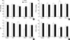
In the LPS+T, the arterial PO2 was lower (p<0.05) and the arterial PCO2 was higher (p<0.05) (Fig. 1A, B) than in the other groups. Administration of the iNOS inhibitor, 1400 W, attenuated the hypoxemia (p<0.05) and the hypercapnia (p<0.05) in the LPS+T group. However, 1400 W administration did not cause such changes in the other groups (Fig. 1A, B).
Arterial pH was lower in the LPS+T group (p<0.05) compared to the other groups, and 1400 W administration attenuated this decrease (Fig. 1C). HCO3- was lower in the LPS+T and LPS+T+P groups compared to the other groups (p<0.05), and 1400 W administration did not affect HCO3- levels (Fig. 1D).

Airway pressures and respiratory compliance

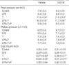
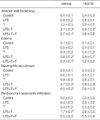
At the end of the experiment, peak airway pressure was higher and quasi-static compliance was lower in the groups with low FRC (i.e., T and LPS+T) compared with the other groups (Table 1). The plateau pressure was higher in the LPS+T group compared with the control or LPS groups (Table 1). The administration of 1400 W did not affect airway pressure or compliance.

Hemodynamics

There was no significant difference in the mean arterial pressure (MAP) between the groups (Fig. 2A), and 1400 W administration did not affect MAP (Fig. 2A).
Although cardiac output appeared to be lower in the LPS+T group compared with the thoracotomy only group (T) (Fig. 2B), this difference did not reach statistical significance (p=0.08). The 1400 W administration had no affect on cardiac output (Fig. 2B).

Histology

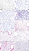
The lungs of the LPS+T group showed marked inflammatory cell accumulation in the interstitium, predominantly comprising of neutrophils and mononuclear cells (Fig. 3D). This group also showed substantial alveolar wall thickening and alveolar collapse in patches (Fig. 3D). The recruitment of inflammatory cells in the LPS+T group lungs was attenuated by PEEP application (Table 2). 1400 W administration also attenuated neutrophil infiltration and alveolar wall thickening (Table 2).

Immunohistochemical staining of iNOS protein

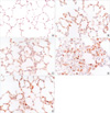
Lung tissue from LPS-treated groups exhibited enhanced immunostaining with the iNOS antibody, predominantly on valveolar mononuclear cells (Fig. 4). These iNOS-stained mononuclear cells accumulated in the interstitium in the LPS+T group, and this accumulation was attenuated by PEEP application (Fig. 4). There was no significant iNOS staining in groups not treated with LPS.

DISCUSSION

The present study shows that atelectasis induced by thoracotomy causes lung injury during mechanical ventilation with conventional tidal volume in endotoxemic rats. The injury was attenuated by either applying PEEP to restore normal FRC or by administration of an iNOS inhibitor. In the injured lung, neutrophils and mononuclear cells accumulated along the alveolar wall, and neutrophils infiltrated the peribronchioles. There was a patchy distribution of inflammation and alveolar collapse in the lungs.
Since a lower tidal volume strategy reduced the mortality rate in patients with acute respiratory distress syndrome (22), many physicians now prefer to use lower tidal volumes when ventilating patients. However, lower tidal volumes may predispose these patients to atelectasis especially if they are associated with high intra-abdominal pressure, pleural effusions, or other conditions (23). Our data suggest that this ventilation at lower lung volumes below FRC may predispose the lungs to further injury. Therefore, restoring the normal FRC by strategies such as higher PEEP may be beneficial in patients prone to atelectasis. Furthermore, increasing the FRC by PEEP increases pulmonary vascular permeability if the same tidal volume is maintained (24). Taken together, maintaining a normal FRC may be important for preventing further lung injury during mechanical ventilation.
The current study found that atelectasis during mechanical ventilation in endotoxemic rats (LPS+T group) caused a significant increase in peribronchial neutrophil infiltration, but significant small airway injury with an increase in airway resistance was not observed compared to rats with atelectasis without endotoxemia. This result is different from the results of the ex vivo saline-lavaged non-perfused rat model that showed significant small airway and alveolar duct injury (25). The main reason for the difference may be that since surfactant is distributed in the small airways as well as alveoli (26, 27), surfactant depletion or inactivation may have contributed to the injury to the small airways seen in a saline-lavaged model emphasizing different lung injury mechanisms of these diverse lung injury models.
Neutrophils and mononuclear cells may be important contributors in the present atelectasis model. iNOS-expressing mononuclear cells accumulated in the interstitium, and iNOS inhibition attenuated recruitment of both mononuclear cells and neutrophils. The accumulation of mononuclear cells and neutrophils was also abrogated by maintaining a normal FRC via PEEP. Taken together, it appears that deformational injury induced by reduced lung volume as well as iNOS is critical for recruitment of inflammatory cells in the present model. iNOS gene expression and activity are upregulated and contribute to neutrophil infiltration in ventilator-induced lung injury, which is also attenuated by iNOS inhibition (15). Although atelectasis induced by the surfactant deactivation model has shown that lung injury is independent of neutrophils (8), neutrophil-depleted rabbits had less lung injury than non-depleted rabbits during conventional mechanical ventilation (28). It is well established that iNOS is crucial for pulmonary sequestration of neutrophils in sepsis models (29, 30). Sepsis is a common cause of ARDS (31, 32). Therefore, either iNOS inhibition or maintaining normal FRC may help prevent progression of atelectasis-related injury in patients with sepsis.
The present study indicated that maintaining normal FRC may be important for preventing inflammatory cell infiltration into the lungs during endotoxemia. In addition, the interaction of repeated opening and closing of alveoli and LPS was found to increase iNOS expression, and an iNOS inhibitor attenuated accumulation of inflammatory cells in the lungs and restored blood gases.
In conclusion, the results of this study suggest that atelectasis induced by thoracotomy causes lung injury during mechanical ventilation in endotoxemic rats through iNOS expression.

Figures and Tables

Fig. 1
Blood gas analysis at the end of the experiment. (A) PO2, (B) PCO2, (C) pH, (D) HCO3-. LPS plus thoracotomy caused a decrease in pH and PO2, and also an increase in PCO2. These changes were attenuated by treating with 1400 W (n=6 per group).
*p<0.05 vs. all other groups with vehicle; p<0.05 vs. vehicle for the same group; p<0.05 vs. control, LPS, and T groups with vehicle; §p< 0.05 vs. control, LPS, and LPS+T+P with vehicle.
jkms-23-406-g001
Fig. 2
Hemodynamic parameters after 2 hr of mechanical ventilation. (A) Mean arterial pressure, (B) cardiac output. There was no significant difference between groups with or without 1400 W (n=6 per group).
jkms-23-406-g002
Fig. 3
Representative histology findings (hematoxylin and eosin stain, original magnification, ×100). Lungs were removed after two hours of mechanical ventilation. (A) Control, (B) LPS, (C) T, (D) LPS+T, (E) LPS+T+P, and (F) 1400 W+LPS+T. Inflammatory cells including neutrophils were infiltrated in the interstitium of collapsed alveolar walls and the peribronchiolar portion in the LPS+T group (D). The group of iNOS inhibition with 1400 W shows reduced interstitial inflammation and alveolar collapse (F). The other groups show unremarkable histologic changes.
Representative histology findings (hematoxylin and eosin stain, original magnification, ×100). Lungs were removed after two hours of mechanical ventilation. (A) control, (B) LPS, (C) T, (D) LPS+T, (E) LPS+T+P, (F) 1400 W+LPS+T. Inflammatory cells including neutrophils were infiltrated in the interstitium of collapsed alveolar walls and peribronchiolar portion in the LPS+T group (D). Group of iNOS inhibition with 1,400 W shows reduced findings of the interstitial inflammation and alveolar collapse (F). The other groups show unremarkable histologic changes.
jkms-23-406-g003
Fig. 4
Immunostaining for inducible nitric oxide synthase (iNOS) expression (original magnification, ×200). (A) Control, (B) LPS, (C) T, (D) LPS+T, and (E) LPS+T+P. The LPS-treated group shows increased expression for iNOS on the mononuclear cells (B). In the LPS+T group, marked expression for iNOS on the mononuclear cells was present in the thickened interstitial areas (D). Less expression for iNOS was present in the thoracotomy alone (C) and LPS plus thoracotomy with the PEEP group (E).
jkms-23-406-g004
Table 1
Airway pressure and compliance at the end of the experiment
jkms-23-406-i001

Cqs, quasi-static respiratory system compliance with tidal ventilation (tidal volume/plateau pressure-positive end expiratory pressure [PEEP]; LPS, lipopolysaccharide; LPS+T, LPS plus thoracotomy; LPS+T+P, LPS+T+PEEP).

*p<0.05 compared with control, LPS+T+P, and LPS groups in vehicle; p<0.05 compared with control and LPS groups in vehicle; p< 0.05 compared with control in 1400 W; §p<0.05 compared with LPS+ T+P in 1400 W.

Table 2
Histologic grading of alveolar wall thickening, edema, neutrophil recruitment, and the presence of neutrophil infiltration in the peribronchioles
jkms-23-406-i002

*p<0.05 versus the other groups in vehicle; p<0.05 compared with vehicle; p<0.05 compared with LPS+T.

LPS, lipopolysaccharide; LPS+T, LPS plus thoracotomy; LPS+T+P, LPS+T+PEEP.

ACKNOWLEDGMENT

The authors thank Yong-Seok Choi for technical assistance.

References

1. Rehder K, Sessler AD, Marsh HM. General anesthesia and the lung. Am Rev Respir Dis. 1975. 112:541–563.
2. Lindberg P, Gunnarsson L, Tokics L, Secher E, Lundquist H, Brismar B, Hedenstierna G. Atelectasis and lung function in the postoperative period. Acta Anaesthesiol Scand. 1992. 36:546–553.
crossref
3. Reissmann H, Bohm SH, Suarez-Sipmann F, Tusman G, Buschmann C, Maisch S, Pesch T, Thamm O, Plumers C, Schulte am Esch J, Hedenstierna G. Suctioning through a double-lumen endotracheal tube helps to prevent alveolar collapse and to preserve ventilation. Intensive Care Med. 2005. 31:431–440.
crossref
4. Muller JM, Erasmi H, Stelzner M, Zieren U, Pichlmaier H. Surgical therapy of oesophageal carcinoma. Br J Surg. 1990. 77:845–857.
5. Tandon S, Batchelor A, Bullock R, Gascoigne A, Griffin M, Hayes N, Hing J, Shaw I, Warnell I, Baudouin SV. Peri-operative risk factors for acute lung injury after elective oesophagectomy. Br J Anaesth. 2001. 86:633–638.
6. Taskar V, John J, Evander E, Robertson B, Jonson B. Surfactant dysfunction makes lungs vulnerable to repetitive collapse and reexpansion. Am J Respir Crit Care Med. 1997. 155:313–320.
crossref
7. Neumann P, Berglund JE, Mondejar EF, Magnusson A, Hedenstierna G. Effect of different pressure levels on the dynamics of lung collapse and recruitment in oleic-acid-induced lung injury. Am J Respir Crit Care Med. 1998. 158:1636–1643.
crossref
8. Steinberg JM, Schiller HJ, Halter JM, Gatto LA, Lee HM, Pavone LA, Nieman GF. Alveolar instability causes early ventilator-induced lung injury independent of neutrophils. Am J Respir Crit Care Med. 2004. 169:57–63.
crossref
9. Woo SW, Hedley-Whyte J. Macrophage accumulation and pulmonary edema due to thoracotomy and lung over inflation. J Appl Physiol. 1972. 33:14–21.
crossref
10. Tremblay L, Valenza F, Ribeiro SP, Li J, Slutsky AS. Injurious ventilatory strategies increase cytokines and c-fos m-RNA expression in an isolated rat lung model. J Clin Invest. 1997. 99:944–952.
crossref
11. Quinn DA, Moufarrej RK, Volokhov A, Hales CA. Interactions of lung stretch, hyperoxia, and MIP-2 production in ventilator-induced lung injury. J Appl Physiol. 2002. 93:517–525.
12. Gosgnach W, Messika-Zeitoun D, Gonzalez W, Philipe M, Michel JB. Shear stress induces iNOS expression in cultured smooth muscle cells: role of oxidative stress. Am J Physiol Cell Physiol. 2000. 279:C1880–C1888.
crossref
13. Kristof AS, Goldberg P, Laubach V, Hussain SN. Role of inducible nitric oxide synthase in endotoxin-induced acute lung injury. Am J Respir Crit Care Med. 1998. 158:1883–1889.
crossref
14. Frank JA, Pittet JF, Lee H, Godzich M, Matthay MA. High tidal volume ventilation induces NOS2 and impairs cAMP-dependent air space fluid clearance. Am J Physiol Lung Cell Mol Physiol. 2003. 284:L791–L798.
15. Peng X, Abdulnour RE, Sammani S, Ma SF, Han EJ, Hasan EJ, Tuder R, Garcia JG, Hassoun PM. Inducible nitric oxide synthase contributes to ventilator-induced lung injury. Am J Respir Crit Care Med. 2005. 172:470–479.
crossref
16. Lai YL, Hildebrandt J. Respiratory mechanics in the anesthetized rat. J Appl Physiol. 1978. 45:255–260.
crossref
17. Wohl ME, Turner J, Mead J. Static volume-pressure curves of dog lungs-in vivo and in vitro. J Appl Physiol. 1968. 24:348–354.
crossref
18. Garvey EP, Oplinger JA, Furfine ES, Kiff RJ, Laszlo F, Whittle BJ, Knowles RG. 1400 W is a slow, tight binding and highly selective inhibitor of inducible nitric-oxide synthase in vitro and in vivo. J Biol Chem. 1997. 272:4959–4963.
19. Thomsen LL, Scott JM, Topley P, Knowles RG, Keerie AJ, Frend AJ. Selective inhibition of inducible nitric oxide synthase inhibits tumor growth in vivo: studies with 1400 W, a novel inhibitor. Cancer Res. 1997. 57:3300–3304.
20. Krieglstein CF, Cerwinka WH, Laroux FS, Salter JW, Russell JM, Schuermann G, Grisham MB, Ross CR, Granger DN. Regulation of murine intestinal inflammation by reactive metabolites of oxygen and nitrogen: divergent roles of superoxide and nitric oxide. J Exp Med. 2001. 194:1207–1218.
21. van Kaam AH, Lachmann RA, Herting E, De Jaegere A, van Iwaarden F, Noorduyn LA, Kok JH, Haitsma JJ, Lachmann B. Reducing atelectasis attenuates bacterial growth and translocation in experimental pneumonia. Am J Respir Crit Care Med. 2004. 169:1046–1053.
crossref
22. The acute respiratory distress syndrome network. Ventilation with lower tidal volumes as compared with traditional tidal volumes for acute lung injury and the acute respiratory distress syndrome. N Engl J Med. 2000. 342:1301–1308.
23. Wongsurakiat P, Pierson DJ, Rubenfeld GD. Changing pattern of ventilator settings in patients without acute lung injury: changes over 11 years in a single institution. Chest. 2004. 126:1281–1291.
24. Dreyfuss D, Saumon G. Role of tidal volume, FRC, and end-inspiratory volume in the development of pulmonary edema following mechanical ventilation. Am Rev Respir Dis. 1993. 148:1194–1203.
crossref
25. Muscedere JG, Mullen JB, Gan K, Slutsky AS. Tidal ventilation at low airway pressures can augment lung injury. Am J Respir Crit Care Med. 1994. 149:1327–1334.
crossref
26. Enhorning G, Holm BA. Disruption of pulmonary surfactant's ability to maintain openness of a narrow tube. J Appl Physiol. 1993. 74:2922–2927.
crossref
27. Enhorning G, Duffy LC, Welliver RC. Pulmonary surfactant maintains patency of conducting airways in the rat. Am J Respir Crit Care Med. 1995. 151:554–556.
crossref
28. Kawano T, Mori S, Cybulsky M, Burger R, Ballin A, Cutz E, Bryan AC. Effect of granulocyte depletion in a ventilated surfactant-depleted lung. J Appl Physiol. 1987. 62:27–33.
crossref
29. Razavi HM, Wang le F, Weicker S, Rohan M, Law C, McCormack DG, Mehta S. Pulmonary neutrophil infiltration in murine sepsis: role of inducible nitric oxide synthase. Am J Respir Crit Care Med. 2004. 170:227–233.
30. Numata M, Suzuki S, Miyazawa N, Miyashita A, Nagashima Y, Inoue S, Kaneko T, Okubo T. Inhibition of inducible nitric oxide synthase prevents LPS-induced acute lung injury in dogs. J Immunol. 1998. 160:3031–3037.
31. Fowler AA, Hamman RF, Good JT, Benson KN, Baird M, Eberle DJ, Petty TL, Hyers TM. Adult respiratory distress syndrome: risk with common predispositions. Ann Intern Med. 1983. 98:593–597.
crossref
32. Hudson LD, Milberg JA, Anardi D, Maunder RJ. Clinical risks for development of the acute respiratory distress syndrome. Am J Respir Crit Care Med. 1995. 151:293–301.
crossref

Notes

This study was supported in part, by a Bisa Research Grant of Keimyung University in 2003 (W-IC); Shriner Hospital, Boston, No. 8620; HL 039150 (CAH).

TOOLS
ORCID iDs

Jeong Wook Seo
https://orcid.org/http://orcid.org/0000-0003-0242-1805

Similar articles