

 PDF
PDF ePub
ePub Citation
 Print
Print


INTRODUCTION
Nasal dorsal cyst formation is one of the rare and late complications of rhinoplasty. Only several cases have been reported in the literature and most of them concerned mucous cysts (1-3). Although many theories have been suggested for the late cyst formation after rhinoplasty, migration or incorporation of mucosal tissue in the subcutaneous space during the surgical procedure has been accepted as an important mechanism (4-6). Careful dissection and meticulous manipulation of tissue and implant are necessary to prevent the mucosa from being grafted with the implant material (4-6). Rarely, a foreign body type cyst resulting from petroleum jelly impregnated with the packing material has been reported.
The authors report a case of dorsal nasal cyst that was presumed to have a different pathogenesis. We describe this case with a brief review of the literature with an emphasis on its pathogenesis and treatment.
CASE REPORT
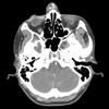
A 58-yr-old woman with a nasal radix mass visited our clinic. Thirty years earlier at a local clinic, she had undergone augmentation rhinoplasty with a material presumed to be silicone. She had had no complication afterwards. However, 5 yr previously, a nasal mass developed that had increased gradually. Physical examination showed a 3.5×2.5 cm round, soft, painless mass on the midline of nasal radix (Fig. 1A, B). The nasal dorsum and the tip were firm on palpation and a small cruciate incision scar was found at the infratip lobule, which was supposed to be the entry of the augmentation material. On computed tomography (CT) scan, a heterogeneous cystic mass was observed at the radix and the cyst was continuous with a homogenous density at the nasal dorsum, which was first thought to be a silicone implant (Fig. 2).
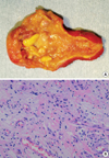
A presurgical diagnosis of nasal radix cyst associated with previous augmentation rhinoplasty was made, and a direct, open approach was chosen to remove the cyst. After a skin incision on the center of the cyst, it was dissected down to the nasal bone. The cyst was connected to the dorsal mass, which were removed together in en-bloc fashion (Fig. 3A). For safe removal of the graft material over the tip area and effective tip-plasty, the tip was exposed with an open approach.
Grossly, the removed cystic mass had a thick, fibrous wall with dirty cheesy material inside. The cystic wall was composed of dense fibrous tissue containing dispersed, microsized amorphous foreign materials (Fig. 3B). Foreign materials were surrounded by foreign body type multinucleated giant cells and induced granulomatous reactions. Infiltration of chronic inflammatory cells was also observed.
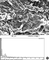
A scanning electron microscopy (SEM) showed a fragment of foreign body (Fig. 4A) and energy dispersive radiography spectroscopy (EDX) showed presence of silicone (Si) in the removed specimen (Fig. 4B). The lesion was diagnosed as a foreign-body type cyst associated with silicone material used for the augmentation rhinoplasty.
The depressed radix, dorsum, and tip after excision of the mass were reconstructed using auricular and septal cartilages. The redundant and thin skin of the radix was trimmed and reinforced with an underlay of the temporalis fascia. A tip-plasty was done using shield and cap grafts. The patient has been followed for 9 months postoperatively with a good aesthetic result and no complication (Fig. 1C, D).
DISCUSSION
Exact pathogenesis of the cyst formation in our case is not clear because no information about the material that had been used for dorsal augmentation was available. However, considering the gross and histological findings as well as the results from SEM-EDX analysis, foreign body inflammatory reaction from silicone material, most possibly a liquid type, which had been injected 30 yr ago, probably caused the cyst.
This reasoning was supported by several facts. First, SEM-EDX analysis of the cyst revealed silicone in the cyst, which meant that a type of silicone material was used for dorsal augmentation. Second, silicone, whether it is a liquid type or implant type, was and still is the most commonly used material for augmentation rhinoplasty in Korea. The patient also remembered that a silicone material was used for her dorsal augmentation. Third, a small, cruciate incision of the infratip lobule was too small to insert any type of hard material including silicone implant. Although calcification and inflammatory cell infiltration around a silicone implant have been reported, total degradation is impossible considering the bio-characteristics of the silicone implant (7, 8).
SEM-EDX used for analyzing the graft material in this case adopted the principles that when the electron generated from the electron microscope collides with the object, a specific radiography characteristic to the object is released from the surface. EDX analysis clarifies the object by detecting this radiography, and this technique is widely used in material engineering and archeology. In our case, a small amount of silicone detected in the specimen led us to believe that the foreign body that caused cyst formation was a type of silicone material.
Silicone implant has been used for rhinoplasty since 1950 and it still remains one of the most widely used implant materials in Asian countries, including Korea (9-11). Injectable liquid silicone is useful in augmenting the chin, cheek, and glabella area (12). It is not recommended for rhinoplasty because the nasal skin is thin and the silicone frequently results in ridging or beading (12). Serious complications such as severe edema or localized discoloration of the injected area has also been reported (13). Only one case of nasal dorsal cyst after augmentation rhinoplasty using silicone implant has been reported in the literature but it's pathologic findings and pathogenesis were not clarified (1).
The mainstay of treating postrhinoplasty nasal dorsal cyst is complete resection and reconstruction. In this case, a direct open approach using the horizontal incision over the cyst center was used given the location and size of the cyst. To expose the tip, an open approach using separate transcolumellar incision was chosen because a previous cruciate incision was located at the infratip lobule and it was too small to expose the nasal tip. In revision rhinoplasties due to complications of the allograft implant, autogenous materials are best to prevent complications. Fortunately, we could augment the depressed dorsum and radix with autogenous septal and auricular cartilages without harvesting the rib cartilage despite the large defect.

 XML Download