

 PDF
PDF ePub
ePub Citation
 Print
Print


INTRODUCTION
A brain abscess is a serious and life-threatening complication of bacterial meningitis. Among the patients with intracranial complications of pneumococcal meningitis in adults, the multiple brain abscesses is a very rare condition (1-3). To the best of our knowledge, the evolution of multiple brain abscesses by Streptococcus pneumoniae has not been reported from serial magnetic resonance (MR) imagings. Here, we report a patient with pneumococcal meningoencephalitis in whom different stages of multiple brain abscesses were documented by serial MR imagings and MR spectroscopy.
CASE REPORT
A 42-yr-old man, who was a chronic alcoholic, was admitted to our hospital presenting with decreased mentality, high fever, and dyspnea. Three days prior to admission, he complained of a headache and shivering. On admission, he had a difficulty in breathing and was unable to converse normally. Vital signs included a body temperature of 38.4℃, pulse rate of 108 beats/min, respiration rate of 22 breaths/min, and blood pressure of 140/103 mmHg. He was unable to open his eyes and made incoherent replies in response to questions. Coarse breathing sounds with rales were auscultated on his left chest. Laboratory findings included: white blood cell 38,500/µL, platelet count 45,000/µL, and blood glucose 166 mg/dL. The cerebrospinal fluid (CSF) analysis included white blood counts of 1,300/µL (predominantly neutrophils; 68%), protein 552 mg/dL, and glucose 1 mg/dL. The CSF stain revealed many Gram-positive cocci. The initial chest radiography revealed a lobar consolidation in his left lower lung field. We empirically started intravenous antibiotics (vancomycin 2 g/day; ceftriaxon 2 g/day; ampicillin 1.5 g/day), to be maintained for 6 weeks, and intravenous dexamethasone 40 mg for initial 4 days.
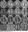
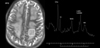
T2-weighted imagings (T2WI) disclosed hyperintense lesions in the bilateral centrum semiovales which could be considered as a hemodynamically internal borderzone area. The dimensions of these lesions were wider than in the initial period. Diffusion-weighted imagings (DWI) showed a high signal intensity in the above regions. Apparent diffusion coefficient (ADC) revealed either several small lesions of decreased ADC, or slightly increased or normal ADC value; these lesions then turned into only hyperintense lesions after 14th hospital day (HD). On gadolium-enhanced T1-weighted imagings (T1E), multiple ring-enhanced lesions with ill-defined rims occurred after HD 7, and the rims became thicker and better-defined even in the ring-enhanced lesion were small (Fig. 1). MR spectroscopy showed elevated peaks of some amino acids, lipid, and lactate on HD 28 (Fig. 2). S. pneumoniae were isolated from the blood and CSF cultures. Three days after admission, the patient showed spontaneous eye opening, but could not raise his left arm and leg in response to painful stimuli. His mentality was slowly improved, and the peripheral leukocytosis and abnormal findings on CSF study were normalized after about HD 14. But even two months after admission, his left extremities were not able to move against gravity in response to painful stimuli.
DISCUSSION
Our patient barely survived and remained with severe motor weakness as a consequence of multiple brain abscesses. Although there are some reports of brain abscesses in patients with pneumococcal meningoencephalitis, this would be the first reported case of serial MR imagings during the evolution of multiple brain abscesses in a patient with lobar pneumonia and meningoencephalitis caused by S. pneumoniae.
According to previous reports, brain abscesses may occur as a result of spread from a contiguous focus or a hematogenous spread from a distant focus, assuming it does not occur as a result of either trauma or neurosurgery (3). Multiple brain lesions in the subcortical borderzone in this case can be considered hematogenous septic embolism because microorganisms are easily deposited in the areas of disrupted perfusion (4).
During the early period (HD 1 to HD 7), the lesions showed hyperintensity on DWI, and hypo-, iso-, or hyperintensity on ADC map. Hence, these findings might depict heterogenous conditions, such as cytotoxic and vasogenic edema. Interestingly, the dimension of the lesions on T2WIs was wider compared to initial periods, even as the size of ring-enhanced regions shrank, and the clinical manifestations and CSF abnormalities were gradually ameliorated. These findings might be obvious evidence of active inflammation working to sequestrate the lesion and protect the surrounding normal brain parenchyma from additional damage, even in the final or capsular stage of a brain abscess (5).
Generally, brain abscesses show ring-shaped enhancement and marked peri-lesional edema, and exhibit restricted water diffusion on MR imaging (6). Additionally, MR spectroscopy findings of brain abscesses may show the presence of acetate, lactate, alanine, succinate, pyruvate, and amino acids because various metabolites are fermented in the central necrotic area. In particular, amino acids are important to discriminate between an abscess and a brain tumor (7).
In conclusion, we described serial brain MR imagings in a patient with penumococcal meningoencephalitis showing not only the evolution of rarely occurring multiple brain abscesses but also the actual evidence of active inflammation even in the late stage of multiple brain abscesses.
 XML Download