

 PDF
PDF ePub
ePub Citation
 Print
Print


INTRODUCTION
Hydroxyurea (HU) is a cytoreductive agent commonly used in the treatment of chronic myeloproliferative disorders. It is usually well tolerated; however, long term HU therapy has been associated with cutaneous side effects such as alopecia, diffuse hyperpigmentation, erythema, skin atrophy and nail changes (1). Poikilodermatous dermatomyositis like skin eruption with atrophy, erythema and scaling have also been reported (2). Leg ulceration following HU therapy is less common. Objective of this paper is to increase the awareness of HU induced leg ulcers which will aid in the early diagnosis and appropriate treatment.
CASE REPORT
Case 1
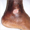
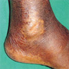
54 yr old male patient with chronic myeliod leulemia (CML) on HU 1.5 g/day since 1996 presented in June 2003 with bilateral perimalleolar ulcers. He had developed ulcers 6 months back, not preceded by trauma. Patient was a known diabetic who was on oral hypoglycemic agents and was switched over to insulin since the onset of ulcers. He was treated with debridement and daily dressing for 6 months but the ulcers did not heal. Examination revealed 1.5×2 cm perimalleolar ulcers on both the lateral malleoli covered by slough with sloping edges, with no evidence of peripheral neuropathy (Fig. 1). The lower limb pulsations were well felt and there were no varicose veins. Investigations including total leukocyte count, fasting and postprandial sugars, RFT, LFT were normal. HU was discontinued and the ulcers healed in 3 months (Fig. 2). Patient was treated with busulphan as he could not afford imatinib.
Case 2
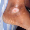
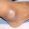
46 yr old CML patient on HU therapy (1.5 g/day) since 2000 presented with bilateral leg ulcers in July 2003. Examination revealed bilateral lateral malleolar ulcers (Fig. 3), and hepatosplenomegaly. There was no evidence of varicose veins or peripheral neuropathy. Local wound treatment did not heal the ulcers. Hydroxyurea was stopped and substituted with imatinib mesylate. The ulcers healed completely in 2 months and the disease was under control with the latter drug (Fig. 4).
Case 3
52 yr old male patient with polycythemia vera on HU 1 g/day, since September 1998 presented to us with painful ulcer over the medial malleolus of left foot of 6 months duration in 2003 with no history of preceding trauma. Past history was significant for a history of stripping of varicose veins in 1992. Examination revealed a 1.5×3 cm ulcer over left medial malleolus covered by slough with sloping edges, bilateral varicose veins and hepatosplenomegaly. Peripheral pulsations were well felt. A differential diagnosis of HU ulcer and venous ulcer were considered. The Doppler study showed normal study of deep venous system of lower limbs with no evidence of saphenofemoral or saphenopopliteal incompetence. There were superficial varicosities along the medial aspect of legs and thighs. The biopsy of the ulcer showed granulation tissue composed of proliferating capillaries lined by plump endothelial cells surrounded by sparse chronic inflammatory infiltrate overlying fibrocollagenous tissue. Surface was covered by cell debris, fibrin and acute inflammatory cells. HU was considered in view of painful non healing nature of the ulcer. The drug was stopped and the ulcer healed promptly in 3 weeks.
Case 4
66 yr old patient with polycythemia vera on HU 1.5 g/day since February 2000 presented in July 2002 with an ulcer on the right lateral malleolus. Local antibiotics, compression stockings and daily wound dressing failed to heal the ulcer. On examination there was an ulcer on the right lateral malleolus with a size of 1.5×3 cm, and hepatosplenomegaly. There were no varicose veins in the lower limbs. HU dose was decreased to 500 mg/day, but the ulcer persisted. Finally HU was stopped and the patient was advised regular phlebotomy. The ulcer healed promptly in 6 weeks.
DISCUSSION
Stahl and Silber first reported HU related skin ulcers in 1985 (3). Frequently such ulcers occur over the malleoli. One explanation for such perimalleolar predilection is the frequently occurring trauma to this site. Histology examination of the edge of the ulcer showed non diagnostic changes. In our case series of 4 patients, non-traumatic perimalleolar ulcers developed while they were on long term treatment with HU for polycythemia vera or CML. The duration of HU therapy ranged from 2.5 yr to 7 yr and the cumulative dose was 1.3 kg to 3.7 kg prior to the ulcer development. Efforts to initiate wound healing before discontinuation of treatment included the use of topical and systemic antibiotic therapy, topical prednisone and topical wound dressings, but with no significant clinical benefit. Our case series indicates that even with meticulous wound care, resolution of the ulcers usually requires discontinuation of HU treatment.
Insidious onset, indolent behaviour and prompt healing after stopping HU suggest that the mechanism for cutaneous toxicity is progressive but reversible cytological damage. HU converts a free radical nitroxide in vivo and inhibits DNA repair (4). Cellular injury accumulates to the extent that repair mechanisms can no longer regenerate viable, normal functioning epidermis, stromal cells or endothelium and tissue damage and ulcers therefore develop (4).
In a review of 14 patients published from the Mayo Clinic, typical site was perimalleolar region with rare sites being the front of the leg, feet and the calf. Multiple ulcers were seen in 9 patients. The ulcers were extremely painful. The average cumulative dose of HU, which the patient received prior to ulcer development, ranged from 1.4 kg to 5.5 kg. The duration of HU treatment ranged from 2 to 15 yr. HU was discontinued in all patients and ulcer healed spontaneously in 12 patients within 2 to 6 months and grafting was done in 2 patients. Two patients developed ulcer after treatment was restarted (5). Montefusco and colleagues described 17 patients who had HU related leg ulcers and found complete resolution (14 patients) or marked improvement (3 patients) after HU therapy was discontinued (6).
Our case series summarized here highlights the association between HU treatment and the development of perimalleolar ulcers in the leg. Our report also confirms the failure of local or systemic antibiotics and the local dressings in curing HU ulcers, and the necessity for discontinuation of the drug and search for alternative therapy once this unusual toxicity develops. We hope that increased awareness of this association will promote early diagnosis and proper management of these ulcers.
 XML Download