

 PDF
PDF ePub
ePub Citation
 Print
Print


INTRODUCTION
Mesenchymal stem cells (MSCs) are cells that have been demonstrated to have the ability to differentiate into distinct mesenchymal tissues such as bones, tendons, muscles, adipose tissues, cartilage and nerve tissues as well as to support hematopoiesis (1-5). MSCs have been labeled in a variety of ways since Dexter named them 'stromal cells' in the 1980's (6). Caplan designated the term mesenchymal stem cells in the 1990's (7), while others still refer to them as marrow stromal cells. The methods used for preparation of these cells have not yet been standardized; the differential potential of MSCs has not yet been fully determined due to the limitations of in vitro expansion. Since Noort et al. reported that MSCs promote engraftment of umbilical cord blood-derived CD34+ cells in non-obese diabetic/severe combined immunodeficiency (NOD/SCID) mice (8, 9) many clinical trials have been pursued to better understand their role in bone marrow transplantation (10). Unlike transplantation of hematopoietic stem cells (HSCs) only, the questions regarding in vitro expansion remain unanswered; in particular, questions regarding the optimal infusion buffer and whether to use thawed cells or fresh detached cells for cotransplantation of MSCs and HSCs remain unanswered. We studied the optimal ratio of HSCs to MSCs to facilitate engraftment, and tried to determine the appropriate ratio for enhancement of HSCs.
MATERIALS AND METHODS
Human CD34+ cells and MSCs isolation
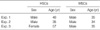
Human CD34+ cells were collected by bone marrow aspiration from 34 to 57 yr-old healthy volunteers (Table 1) under an Institutional Review Board approval protocol of Samsung Medical Center, Korea. CD34+ cells were isolated using the MACS sort system (Miltenyi Biotec GmbH, Germany). The purity of the CD34+-selected cells was more than 90%. MSCs were cultured as previously described (11). We used bone marrow derived MSCs that were not exceeded over 3 passages. MSCs were confirmed to be negative for hematopoietic markers by flow cytometry. For further clarification of MSCs, the following antibodies were used: CD14 (BD-Pharmingen, Palo Alto, CA, U.S.A.), CD29, CD34, CD44, CD45, CD51-61, CD90, CD105, CD106, CD166, HLA-DR, Stro-1, SH2-M2, SH3-M2 and SH4-M2.
Cotransplantation of MSCs and HSCs into NOD/SCID mouse
NOD/SCID mice, purchased from Jackson Laboratories (MA, U.S.A.) were maintained in microisolator cages in specific pathogen free conditions. Eight-week old mice received sublethal total-body irradiation with 300 cGy from 137-Cs source within 24 hr before transplantation. In all experiments, 1×10⁵ bone marrow derived human CD34+ cells were administered to each mouse and MSCs were used concomitantly. Bone marrow derived CD34+ cells and MSCs were injected in a final volume of 500 to 1,000 µL HBSS (Bio-Whittaker, Baltimore, MD, U.S.A.) per mouse and were infused via tail vein injection. Three experiments were performed repeatedly. Serial ratios of bone marrow derived CD34+ cells to MSCs were 1:0, 1:1, 1:2 and 1:4, in the first experiment (4 mice), 1:0, 1:1, 1:2, 1:4 and 1:8 in the second (10 mice) and 1:0, 1:1, 1:4, 1:8 and 1:16 in the third (15 mice).
Analysis of human CD34+ cell engraftment
Mice were sacrificed 4 weeks posttransplant and cells from the bone marrow were analyzed by flow cytometry. Cells were then incubated with PE-mouse anti-human CD45 (BD-Pharmingen, Palo Alto, CA, U.S.A.) for 30 min at 4℃, washed, resuspended in 1% paraformaldehyde in phosphate-buffered saline, and analyzed on a flow cytometry (FACS Vantage, Becton Dickinson, Palo Alto, CA, U.S.A.).
RESULTS
The immunophenotye of bone marrow derived HSCs
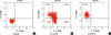
We isolated CD34+ HSCs from bone marrow using CD34+ cell directed kit (Miltenyi Biotec GmbH, Germany), and at the same time, isolated MSCs from CD34- population. The CD34+ cells infused had purities over 90% and CD34+/CD38- cells were in the range of 30±5-40±5% (data not shown) (Fig. 1).
The immunophenotyes of bone marrow derived MSCs
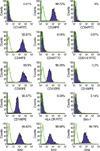
FACS results showed that CD29, CD44, CD90, CD105, CD166, SH2, SH3, and SH4 were expressed in more than 90% of MSCs. Typical hematopoietic antigens (CD14, CD34 and CD45) as well as CD51-61, CD106 and HLA-DR known to be negative markers for MSCs (2, 12, 13) were hardly expressed. On the other hand, Stro-1, known to be related with homing to bone marrow (14), was expressed in 3.14% of MSCs (Fig. 2).
Effect of MSCs on engraftment according to cell dose
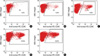
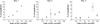
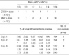
We observed that the engraftment efficiency after cotransplantation was improved as the MSCs dose was increased up to a certain level in all three independent experiments. The engraftment fold-increase of the 1:4 group was 3.99, 19.78 and 6.03 compared to the HSCs only group in experiment 1, 2 and 3, respectively (Table 2). The percent donor chimerism of the 1:16 group was inferior to that of the 1:8 ratio group (Fig. 3). The 1:4 and 1:8 ratio groups were associated with a higher percent donor chimerism than any other group studied (Fig. 4).
DISCUSSION
MSCs have recently been used experimentally in clinical trials of cardiovascular repair (15, 16), treatment of lung fibrosis (17), spinal cord injury (18) and bone and cartilage repair (4, 19) as a stem cell source. MSCs have been successfully used for therapeutic application when they are derived from bone marrow (20).
Generally, an accepted range of HSC dose for engraftment after bone marrow transplantation is 3-4×10⁶ CD34+ cells/kg of recipient body weight for humans. Considering that a mouse weighs about 30 g on average, we postulated that 9-12×104 CD34+ cells should be a reasonable dose range for a mouse, and therefore infused 1×10⁵ CD34+ cells.
Usually, the fluorescence intensity of CD34+ cells when conjugated with FITC is weaker than that when conjugated with PE using flow cytometry. Therefore, it is a matter of course that the percentage of CD34+ fraction may is different to some extent according to the conjugated materials even though the testing samples are exactly the same. Authors usually evaluate the purity of overall CD34+ cell fraction stained with PE according to the manufacturer's protocol of CD34 sort kit (Miltenyi Biotec, Germany). Fig. 1B is intended to show the percentage of CD34+/CD38- fraction which is known to be more primitive and have higher engraftment potential than CD34+/CD38+ fraction using PE-conjugated CD38 and FITC-conjugated CD34.
At present we do not know how long MSCs will maintain innate characteristics including the ability to differentiate and cytokine production; nor do we know what the best composition culture media should contain for maintaining MSCs. We used early MSCs not exceeding 3 passages based on the assumption that early passage cells would be more likely to have the innate characteristics of MSCs.
Cotransplantation of HSCs and MSCs enhanced engraftment as the dose of the MSCs were increased up until a 1:8 ratio. The percent donor chimerism in the 1:16 group, however, was lower than that of the 1:8 group even though it was still higher than the HSCs only group as shown in the experiment 3. These findings suggest that HSCs and MSCs have a threshold ratio of HSCs to MSCs for enhancing engraftment when cotransplanted into a NOD/SCID mouse. Our findings suggest that the optimal ratio of HSCs to MSCs in cotransplantation might be between 1:8 and 1:16 and we are conducting further experiments using these ratios. We confirmed MSCs engraftment enhancing ability in 3 experiments. Each experiment was performed with bone marrow-derived MSCs obtained from a different volunteer donor, and showed different enhancing abilities. For example, the mean fold-increase of the 1:4 ratio group was 3.99 compared with the HSCs only group in the experiment 1, while it was 7.21 in the experiment 2, and 1.21 in the experiment 3. The CD34+ cells were also derived from different donors; however, it is unlikely that the quality of MSCs is the sole determinant of engraftment efficiency. It is clear that the CD34+ cell dose has been shown to be strongly associated with engraftment kinetics regardless of individual differences (21, 22).
In addition, the proliferation rate and morphology of the 3 MSCs experiments differed when observed during cell expansion before cotransplantation (data not shown). Therefore, our findings suggest that the engraftment enhancing ability of MSCs might have considerable difference according to the MSC donor.
Our study demonstrated that MSCs could enhance engraftment in a dose-dependent manner up to a certain level in NOD/SCID mice. Future studies should help elucidate further mechanisms associated with engraftment such as secretion of specific MSC-cytokines that help enhance engraftment. For clinical application to human patients characteristics of MSCs including immunosuppression and HLA restriction require further explanation.
 XML Download