

 PDF
PDF ePub
ePub Citation
 Print
Print


INTRODUCTION
Vancomycin-resistant enterococci (VRE) have recently emerged as important nosocomial pathogens that mainly affect patients with severe underlying diseases (1, 2). VRE was first reported In Korea in 1992. Since then, however, the incidence of VRE was low until 2000 when an increase in VRE infections began to be reported in many hospitals (3-6). A nationwide survey suggested that the main mode of transmission was interhospital spread (4). Nowadays it is clear that VRE is a major newly established pathogen in Korean hospitals.
At the end of 2000, we recognized a sudden increase in the frequency of hemato-oncological patients with VRE bacteremia (8 patients) in our institution compared with previous years when there was 1 bacteremia in 1997 and 2 each in 1998 and 1999. This trend continued in 2001 in which there were 11 hemato-oncological patients with VRE bacteremia.
We herein conducted a retrospective analysis of the clinical and molecular epidemiologic characteristics of the VRE bacteremic patients who received hematopoietic stem cell transplantation (HSCT) or cytotoxic chemotherapy between January 2000 and December 2001.
MATERIALS AND METHODS
Setting and patients
The Catholic HSCT Center belongs to St. Mary's Hospital, a 600-bed, university-affiliated, tertiary care teaching hospital. The HSCT center deals with more than 250 HSCT per year, which represents more than half of the annual HSCT cases in Korea. It follows that the analysis of clinical data for patients at our institution should provide reliable information about overall trends in hemato-oncological patients in Korea.
We reviewed retrospectively the medical records of all 19 VRE bacteremic patients over a 2-yr period (January 2000 through December 2001) in the Catholic HSCT Center affiliated with the Catholic University of Korea, College of Medicine.
Fever was defined as a single oral temperature of ≥38.3℃ or a temperature of ≥38.0℃ for ≥1 hr. Neutropenia was defined as a neutrophil count of less than 500 cells/µL, or a count of less than 1,000 cells/µL with a predicted decrease to less than 500 cells/µL.
The term clinically significant VRE bacteremia was defined as at least two blood cultures positive for vancomycin-resistant Enterococcus faecium or E. faecalis (7).
An empirical antibiotic regimen was given to febrile patients according to the guidelines of the Infectious Diseases Society of America (8).
Identification of isolates, and testing of antibiotic susceptibility
Identification was performed with a conventional Microscan panel (Dade International, West Sacramento, CA, U.S.A.). VRE isolates were tested for antimicrobial susceptibility by the agar dilution method and the results were interpreted according to the Natinal Committee for Clinical Laboratory Standards 2002 breakpoints (9). Enterococcus faecalis ATCC 29212 and Staphylococcus aureus ATCC 29213 were used for quality control.
Genotyping
Vancomycin resistance genotypes (vanA, vanB, vanC1, vanC2/C3) were tested by amplifying the respective genes by PCR (10), with some modifications. Primers for vanA were 5'-GGGAAAACGACAATTGC-3' (175-191) and 5'-ATTGCCGGCGTAACATG-3' (891-907); for vanB, 5'-ATGGGAAGCCGATAGTC-3' (173-189) and 5'-CCAGCTCCTTGCTTTAG-3' (791-807); for vanC1, 5'-GGTATCAAGGAAACCTC-3' (246-272) and 5'-TCGATACTACCGCCTTC-3' (1051-1067), and for vanC2/C3, 5'-CTCCTACGATTCTCTTG-3' (455-486) and 5'-GAATTTCCAGAACGAGC-3' (869-885). Strains E. faecium BM4147 with vanA and ddIE. faecium, E. faecalis V583 (vanB, ddIE. faecalis), E. gallinarum BM 41745 (vanC1), and E. casseliflavus ATCC 24788 (vanC2/C3) served as positive controls.
Molecular epidemiological typing
Fourteen of the 19 VRE isolates were typed by pulsed field gel electrophoresis (PFGE) with SmaI as restriction enzyme, as previously described in detail (6). In addition, we obtained VRE isolates from specimens other than blood (urine and stool) that were available from eight patients without bacteremia (July 2001 through December 2001). These additional non-bacteremic (NB) isolates were also subjected to PFGE to investigate whether they might involve strains identical to or closely related (2-3 fragment differences) to strains in the patients with bacteremia.
PFGE was performed with a CHEF-DR II apparatus (Bio-Rad Korea, Seoul, Korea). The conditions were: initial switch time 0.5 sec, run time 20 hr, final switch time 40 sec. After ethidium bromide staining, patterns were compared and anayzed with Uniband/Map (version 99; UVItech, Cambridge, U.K.).
Data collection and analysis
We compared VRE bacteremic patients with a group of hematological patients with vancomycin-susceptible E. faecium (VSE) bacteremia (n=8) arising during the same period.
Baseline information included age, sex, and underlying disease. A simplified acute physiologic score (SAPS II) was assigned to all patients at the onset of bacteremia (11). We also followed up the patients' record until January 2002, to permit a survival analysis. The starting point of survivial was the date when bacteremia was detected.
Statistical analyses were carried out with SPSS 10.0 (SPSS Korea, Seoul, Korea). If the data were in the category of continous variables, t-test was performed. In case of data with ordinal or nominal scale, nonparametric tests such as chisquare, Mann-Whitney test, etc were done. P values <0.05 were considered statistically significant.
RESULTS
Clinical characteristics of patients with VRE bacteremia
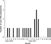
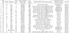
Table 1 gives the characteristics of the patients with VRE bacteremia. There were 8 cases of VRE bacteremia in the year 2000, and 11 cases in 2001 (Fig. 1). Eighteen of the 19 patients (94.7%) received broad-spectrum antibiotics prior to detection of the bacteremia. Only eight patients with VRE were able to receive quinupristin/dalfopristin (Q-D) because this was only formally available in our institution from 2001. The other 11 patients received high-dose ampicillin/sulbactam plus gentamicin, or imipenem monotherapy. Four of the 8 patients receiving Q-D died, compared with all the patients who did not receive Q-D (11/11).
Microbiological characteristics of patients with VRE bacteremia
All 19 VRE isolates were identified as E. faecium. All were also vanA genotype. Their antimicrobial susceptibility profiles showed high-level gentamicin resistance (100%), whereas only 14.3% had high-level resistance to streptomycin. None of them were resistant to Q-D or linezolid.
Comparison of the clinical characteristics with VSE-bacteremia
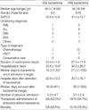
Table 2 compares the demographic characteristics of patients with VRE versus those with VSE bacteremia. Simplified acute physiology score (SAPS) II were similar in both groups (42.6±12.8 in VSE vs. 41.4±13.7 in VRE, p>0.05, respectively), indicating that initial disease severity at the time of bacteremia did not differ significantly. Age, gender ratio, and other demographic characteristics were also similar.
The VSE group had a lower median number of days to bacteremia and fewer days of hospitalization than the VRE group, but the mortality of the two groups did not differ significantly (62.5% for VSE vs. 78.9% for VRE, p>0.05, respectively). More patients with VRE received antibiotics before bacteremia than those with VSE (94.7% vs. 62.5%, p<0.05, respectively). The median days survived for VSE was not statistically different from that for VRE as shown in Table 2 (log rank 1.55, p=0.2130).
Characteristics of patients with VRE bacteremia who survived and of those who died
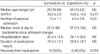
Table 3 compares patients with VRE bacteremia who survived (n=6) with those who died (n=15). Age, number of previous admissions, days to detection, total days of hospitalization, and neutropenic days did not differ significantly between the two groups. But SAPS II was significantly higher in those that died than in those that survived. While all the patients who survived recovered from the neutropenia, 40% of those who died did not.
Molecular epidemiological characteristics of patients with VRE bacteremia
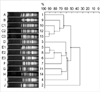
As illustrated in Fig. 1, the occurrence of VRE bacteremia appeared initially to be sporadic. However, its frequency began to increase from April 2001 and reached its peak through June 2001, which suggested us that there might have been a cluster of outbreaks in our institution. We therefore performed PFGE in order to determine whether the VRE bacteremias from 2000 to 2001 were clonally related.
Most of the PFGE fingerprint patterns differed from one another, but there were two very small clusters of clones (C1-3 and E1-3, in Fig. 2 and Table 1). C1 was isolated on 9 May 2001, and C2 appeared about one month later (15 June 2001). As already suggested in Fig. 1, an outbreak did occur during the period May through June 2002, but it was very small (a mini-outbreak). Another closely related strain (C3) emerged 5 months later (22 November 2001) in a room adjacent to where C2 had appeared (room numbers 1308 and 1309, respectively, Table 1). There also occurred another mini-outbreak from December 2000 (E1) through January 2001 (E3). Three months later, a closely-related strain (E2) emerged in the same ward as E1. However the PFGE type E strains did not appear again (up to the end of 2001).
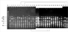
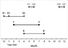
After examining the clonality of VRE bacteremia, we performed additional PFGE analyses, this time of NB isolates, in order to search for a possible source of transmission from patients without bacteremia. As shown in Fig. 3, we found three NB strains which were clonally related to those from patients with VRE bacteremia. The urine isolate (25 July 2001) from patient NB8 (lane 31 in Fig. 3) was virtually identical to PFGE type F which was isolated on 9 February 2001. The stool isolate (30 August 2001) from patient NB7 (lane 30) was closely related to PFGE type B (19 April 2001, lane 9), and the urine and stool isolates (28 December 2001) from patient NB3 (lane 21 and 22) were probably related to PFGE type C2. These patterns are summarized in Fig. 4.
The strains in lane 15 (blood) and 16 (stool) were from the same patient as lane 13 (C3), but the PFGE patterns of the blood and stool isolates differed from each other.
DISCUSSION
In this study, all of 19 isolates were E. faecium of vanA genotype; other vancomycin-resistant genotypes such as vanB or vanC were not found. This feature is consistent with other instances of VRE in Korea. In a Korean tertiary care hospital with 1,550 beds all of 330 VRE isolates were vanA (6). It follows that vanA is the major genotype in Korea although a case of vanB has also been reported (12). The 5th Korean Nationwide Surveillance of Antimicrobial Resistance also revealed an increase in vancomycin-resistant E. faecium in the period 1997-2001 (3).
We used patients with VSE for comparison with the VRE group, as we thought that a control group that simply did not have VRE bacteremia would not be suitable because it would differ in many respects: disease severity as well as clinical characteristics would be quite different from those with VRE, and it would be hard to adjust for these variables. On the other hand, the initial severity of disease as represented by SAPS II was not significantly different in VSE group from that of the VRE bacteremia group. The well-established risk factors for VRE acquisition are neutropenia, prolonged hospitalization, prior use of broad-spectrum antibiotics, and hypoalbuminemia (13-17). These factors, with the exception of days of hospitalization, did not differ significantly between our VRE and VSE groups. However, as the number of patients with VSE (n=8) was very small, and less than half of those with VRE (n=19), we think the variables predictive of the acquisition of VRE should be evaluated in a larger study.
The influence of vancomycin-resistance on mortality is controversial. Some authors have insisted on its importance (18-20), whereas others are of the contrary opinion (21, 22). Since the mortality of our patients with VSE did not differ from those with VRE, it is unlikely that vancomycin-resistance itself affects the prognosis. Our findings suggest that enterococcal infection itself, or its degree of virulence, rather than vancomycin-resistance, is a determinant of prognosis.
Several authors have suggested that Q-D and linezolid would both be effective in treating VRE (23-27). In our study, the patients who received Q-D appeared to do better than those receiving other antimicrobials, though the number of cases (n=8) was too small to be sure.
Among the 19 patients with VRE bacteremia, expired patients had significantly higher SAPS II values than those who survived, and 60% of the latter did not recover from neutropenia. Although further cases are needed to establish the predictors of outcome in VRE bacteremia, we think that initial disease severity, and host immunity as reflected in neutropenia, could be major determinants.
Since its first isolation in 1992, VRE seemed to be rare until 1997 after which an abrupt increase in VRE infections was reported by many hospitals in Korea from 1998 onwards (3-6). One study of VRE infection in a Korean tertiary teaching hospital revealed that the increase in VRE was probably due to the increased use of oral vancomycin to treat Clostridium difficile infections, and clonal spread (6). Another nationwide study in Korea also suggested that interhospital spread contributed to the rapid increase of VRE in many hospitals (4). The findings of our study suggested that spread by a few clones among heterogenous ones may have contributed to the increase in VRE bacteremia. However, it could not be the only mechanism because the PFGE analysis demonstrated a high degree of diversity among the isolates, and bacteremia of endogenous origin could not be excluded. As reported earlier, the hemato-oncological disease itself has been recognized as an important risk factor for the acquisition of VRE (12, 28, 29). In addition to the lack of host immunity, breakdown of the gastrointestinal mucosal barrier and subsequent translocation of bowel flora could play a major role in the acquisition of VRE bacteremia (30-32). In other words, endogenous infection by translocation as well as exogenous transmission from other patients (33, 34) or from fomites (35) could have been another major source of VRE bacteremia in our patients. However, there was also a perplexing but nevertheless important observation arguing against the endogenous route. As illustrated in Fig. 3, blood and stool isolates from one bacteremic patient yielded completely different DNA band patterns (lanes 15 and 16, respectively). If the patient's bacteremia was from her own bowel flora, the DNA patterns of blood and stool isolates should have been identical or at least closely-related. This finding suggested that the VRE, at least in this patient, was not of endogenous origin but came from someone or somewhere else. However, the possibility that the patient may be colonized with more than one strain or that there was transfer of the vanA gene could not be excluded.
While there have been several reports of a single clone dominating a VRE outbreak (29, 36, 37), many molecular epidemiologic studies have shown that most of the VRE in a given hospital is clonally diverse (14, 39-42). In our case, the VRE of our bacteremic patients was clonally diverse with two small clusters of mini-outbreaks (E1-3 and C1-3 in Fig. 2), which suggested that VRE already became endemic in our institution. We also found three strains from non-bacteremic patients were clonally related to those in bacteremic patients (PFGE types B, C2, and F), and each of these strains appeared sporadically at intervals of 4-6 months. Whereas strains of type E (found only in bacteremic patients), B and F did not appear again after August 2001, type C strains continued to emerge up to the end of the study. Based on these findings, we assume that there were multiple clones of VRE coexisting with at least two primary clones of sporadic patterns in our unit, as also postulated in another instance (43). Though both exogenous and endogenous modes of infection could be major causes of bacteremia in our patients, the evidence for mini-outbreaks in the midst of clonal diversity strongly suggests that strict precautions are crucial to prevent nosocomial transmission of VRE (44, 45).
In summary, patients with VRE (all were vanA E. faecium) bacteremia showed heterogenous molecular epidemiologic pattern with some small clusters, but vancomycin-resistance did not seem to be a prognostic factor because of the similar mortality in the VSE group. We consider that greater attention should be paid to the prevention and management of VRE bacteremia because it is now one of the important established pathogens in hemato-oncological patients in Korean hospitals.
 XML Download