

 PDF
PDF ePub
ePub Citation
 Print
Print


INTRODUCTION
Periarticular calcification, characterized by pathological or radiographical evidence that it calcificates around articulations, may be located in cartilage, synovium, capsule, tendons, bursa, ligaments, soft tissue, and vessels (1). Acute calcific periarthritis (ACP) is an acute periarticular inflammation associated with juxtaarticular deposits of calcium hydroxyapatite. Although this entity has been involved in body sites such as shoulder, hip, knee, and elbow, there has been a relative paucity of ACP involving the joints of the fingers (2, 3). Here we report a rare case of ACP in a professional golfer, who continuously practices his swing, caused by repeated minor traumas at the injury site with a review of literatures.
CASE REPORT
A 22-yr-old man visited the Department of Orthopedic Surgery due to painful swelling on the volar surface of the proximal interphalangeal (PIP) joint of the 4th finger that had aggravated for the last 2 weeks. According to the personal history, he is a professional golfer and was facing a professional tour game coming soon. So he started intensive practicing 4 weeks before his visit to our department. On physical examination, he had a swollen, reddened finger which was tender over the PIP joint of the 4th finger. There was no obvious nodule or fluctuant mass. The range of motion (ROM) at PIP joint showed active motion of 10-30° and passive motion of 0-80°. Active and passive ROM at metacarpophalangeal (MP) joint showed 0-80° and 0-90°, respectively. Active and passive ROM at distal interphalangeal (DIP) joint showed 0-80° and 0-90°, respectively.
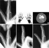
On oblique view of the initial plain radiographs (Fig. 1A), a dense calcified lesion with a triangular shape measuring 5×3 mm was found, and this lesion was located around the 4th PIP joint; however, there was no evidence of joint space narrowing or cortical lesion. This calcification was not identified on standard views. The radiographic presumptive diagnoses included ACP, heterotropic ossification, and an undetermined mass. Three-phasic bone scan showed a hot uptake on the 4th PIP joint (Fig. 1B), and CT demonstrated a triangular calcification at the PIP joint (Fig. 1C). Based on these findings, a diagnosis of ACP was made, and the patient was managed conservatively by ibuprofen 800 mg b.i.d, ice compression and short arm splint immobilization. Two weeks later this calcification was gradually absorbed, the symptom was relieved, and the limitation in ROM was improved (Fig. 1D). One month later the area between PIP joint of the left fourth finger showed only a small, residual calcification (Fig. 1E). Final plain radiograms taken three months after presentation revealed nearly complete resolution of the calcification (Fig. 1F). On physical examination, he showed normal ROM at PIP, DIP, MP joints and the previous symptoms, such as swelling and redness, had completely disappeared. Either discomfort or pain was not felt by the patient during his normal activities, though he felt a mild discomfort while playing golf. Three months later he expenenced no recurrence even after the golf tournament.
DISCUSSION
Periarticular crystal deposition has been described by a variety of terms, such as calcareous tendonitis and bursitis, periarthritis calcarea, calcific tendonitis, peritendinitis and bursitis, and hydroxyapatite rheumatism (1). These deposits are frequently monoarticular in distribution (1), and the shoulder is the most commonly affected site, followed by the hip, knee, elbow, wrist and ankle joints. Finger joints are less commonly involved, and the metacarpophalangeal joint is the most frequently affected site followed by the PIP joint (3). In our case, acute calcification was involved in PIP joint. According to the Carrol et al.'s report (4), this condition occurs in both women and men, with an average age of 45 yr old. Yosipovitch and Yosipovitch (5) reported that it occurs in women far more frequently than in men, between 40 and 70 yrs. In our case, the patient was a man and was in his early 20's, which is different from the previous reports.
The pathologic findings of this entity include calcific material formed into psammoma-like bodies, together with exudates, including many neutrophil leukocytes. Electron microscopy showed the calcific material was composed of hydroxyapatite crystals (6, 7).
Acute symptoms of this condition include, as in our case, pain, swelling, erythema and restriction of motion around the joint involved (1, 3). Chronic symptoms may be present and include mild, non-incapacitating pain and tenderness (1). Carroll et al. (4) reported a history of local trauma in one-third of patients. Yosipovitch and Yosipovitch (5) reported that the hand was constantly exposed to repetitive microtraumas, especially in manual labor. On the other hand, Kim et al. (8) reported that it frequently occurs in association with systemic diseases, such as thyroidism, rheumatoid arthritis, diabetes mellitus, gout and pseudogout. In our case, his job was professional golfer and after intensive practicing, periarticular symptoms such as pain, swelling, and erythema occurred. Therefore, we thought that repeated minor traumas of the joint would be the cause of periarticular calcification.
The radiographic changes in patients with periarticular calcifications show varying patterns; the deposits may remain static for a long period, enlarge and change shape, or diminish in size and disappear (3). In our case, after medication and immobilization this calcification was gradually absorbed. Follow-up plain radiograph after three months showed a subtle calcific deposit and he had no symptom in the joint.
ACP is self-limiting and usually resolves in 3 to 4 weeks even if not treated (9). Recurrences are uncommon. Treatment for ACP involves conservative measures such as splinting and nonsteroidal anti-inflammatory drugs.
In this case, ACP occurred in the PIP joint of a professional golfer with symptoms such as pain, swelling, and erythema. Repeated minor traumas in the affected site was considened to be the cause. Since this entity is self-limiting, correct diagnosis is important to avoid unnecessary tests and surgery.
 XML Download