

 PDF
PDF ePub
ePub Citation
 Print
Print


INTRODUCTION
Choledochal cysts, although rare in adulthood, are now correctly diagnosed more frequently due to recent improvement in biliary imaging techniques (1). There have been numerous descriptions and classifications based on the location and anatomical structure of choledochal cyst. Among these, the most practical classification is proposed by Alonso-Lej et al. (2), which is modified by Todani et al. (3) in 1977. The major point of this classification is that the gallbladder is usually not distended and arises from a small cystic duct, usually not at the upper end of the cyst (4). Initial reports suggested that the following treatments provided satisfactory results: T-tube choledochocystomy with internal or external drainage, cystojejunostomy, modified cystojejunostomy with T-tube external drainage, cyst excision and hepaticoduodenostomy, cystoduodenostomy, jejunal segment interposition with cystoduodenostomy, and jejunal segment interposition with hepaticojejunostomy. However, it is now recognized that these procedures are associated with an unacceptably high rate of late complications. Late complications of internal cyst drainage may result in a high rate of cholangiocarcinoma arising from the cyst (1), as well as the development of cholangitis and biliary stone disease (1, 5-8). Surgical excision of cyst and hepaticojejunostomy are now considered as the treatment of choice for choledochal cysts (1, 5-8). In this paper, we describe our experience with 72 patients with choledochal cysts.
MATERIALS AND METHODS
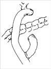
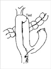
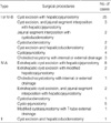
Seventy-two patients (59 females and 13 males) with ages ranging from 15 to 63 (mean age=31) yr were treated for choledochal cyst by the Department of Surgery, the second Affiliated Hospital of Harbin Medical University, between January 1, 1985 and December 31, 2002. The medical records of all 72 patients were reviewed. Choledochal cysts were classified according to Alonso-Lej et al. (2) and Todani's modified classification system (2, 3). Follow-ups were done on all of the patients until December 31, 2002. We analyzed the clinical outcome in patients who underwent the following treatments: cyst excision with hepatojejunostomy (Fig. 1), cyst excision with modified hepatojejunostomy (Fig. 2), and non-cyst excision and without hepatojejunostomy.
RESULTS
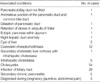
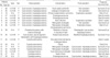
A total of 72 adult patients were identified with choledochal cysts. There were 49 (68.1%) Type I or Type IV-B, one (1.3%) Type II, 21 (29.3%) Type IV-A, and one (1.3%) Type V choledochal cysts. Among the patients, the most common symptom was abdominal pain, which occurred in 64 of the 72 patients (88.9%). Jaundice was present in 40 patients (55.6%), and abdominal mass was present in 21 patients (29.2%). Only eight patients (11.1%) presented with the classic triad of abdominal pain, jaundice, and abdominal mass. The biochemical measurement showed anomalies in AST, which appeared in 22 of the 72 patients (30.6%), ALT in 48.6%, γ-GT in 31.9%, and total bilirubin in 34.7% of all the patients. One or more of the following test procedures established the diagnosis of choledochal cyst: ultrasonography (US) (n=49), magnetic resonance imaging (MRI) (n=4), computed tomography (CT) (n=13) and endoscopic retrograde cholangiopancreatography (ERCP) (n=14). The choledochal cysts were associated with other diseases including the pancreaticobiliary duct not filled in one case, anomalous junction of the pancreatic duct and common bile duct in one case, dilatation of pancreatic duct in one case, retention of stones in ampulla of Vater in one case, ectopic pancreas within jejunum in one case, right hepatic duct anomaly in one case, cyst of liver in two cases, coexistent choledocholithiasis in 12 cases, secondary cholestatic liver cirrhosis, which occurred in one of the 5 cases of intrahepatic cholestasia, cholecystitis in 54 cases, infection of biliary tract in 29 cases and secondary chronic pancreatitis in 5 cases. Women with choledochal cyst were diagnosed during pregnancy (jaundice, abdominal pain) in 3 cases (Table 1).
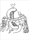
The definite surgical procedures were cyst excision with hepaticojejunostomy in 25 cases for type I or type IV-B and complete excision of extrahepatic choledochal cyst from the hepatic hilum with hepaticojejunostomy in 8 cases for type IV-A, extrahepatic cyst excision with modified hepaticojejunostomy in 2 cases for type IV-A, choledochocystomy with internal or external drainage in 3 cases for type I or type IV-B and 1 case in type IV-A, cyst excision and the biliary tract reconstruction by jejunal segment interposition with hepaticojejunostomy (Fig. 3) in 3 cases for type I or type IV-B and extrahepatic cyst excision and the biliary tract reconstruction by jejunal segment interposition with hepaticojejunostomy in 2 cases for type IV-A, the biliary tract reconstruction by jejunal segment interposition with cystoduodenostomy in 2 cases for type I or type IV-B, cystoduodenostomy in 2 cases for type I or type IV-B and 1 case in type IV-A, cyst excision and hepaticoduodenostomy in 2 cases for type I or type IV-B and 1 case for type II, cystojejunostomy in 4 cases for type I or type IV-B and 5 cases in type IV-A, modified cystojejunostomy with T-tube external drainage for 1 case in type IV-A (Table 2). 27 out of 62 with received operation cases were performed non-cyst excision with or without hepaticojejunostomy in type I, II, IV-A, IV-B. Because of choledochal cyst adhered with surrounding tissue extensively, we can not dissect choledochal cyst from the round ligament in 27 cases. Ten out of the 72 patients did not receive operation.
The early postoperative morbidity and mortality rates were 16.1% (9/62) and 6.5% (4/62) respectively. The complication rate related to the surgical procedure was 30.6% (19/62). The incidence of carcinoma was 6.9% (5/72), while the incidence of cholangiocarcinoma with non-cyst excision or non-operated congenital choledochal cyst was 10.8% (4/37). Early postoperative complications included wound infection, septic shock after cystojejunostomy in one case, acute septic peritonitis after cyst excision with hepatojejunostomy in one case, abdominal pain, pancreatic ductal dilatation with retrograde cholangitis after cyst excision with hepatojejunostomy in one case, which was resolved after a few days with conservative antibiotics treatment.
Two cases required reoperative revision in the early postoperative period due to anastomotic leakage after cyst excision with Roux-en-Y hepaticojejunostomy. Unfortunately, one patient died of brain infection within one week and the other suffered from anastomotic leakage and intra-abdominal hematoma formation after cystoduodenostomy and eventually died of multiorgan failure within one week. During the immediate postoperative period, one patient with fistula formation after cystoduodenostomy died of hepatic failure within 72 hr. In two cases, a 44 yr old male patient and a 37 yr old female, both with type I or type IV-B, were admitted to the hospital with (postoperative) diagnosis of well-differentiated cholangiocarcinoma. Within sixth month following cyst excision with hepaticojejunostomy, both patients died. For one pregnant 27 yr old (type IV-A), a Roux-en-Y cystojejunostomy with T-tube external drainage was performed. However, primigravida presented at 20 weeks' gestation, and the patient died of septic shock and multiorgan failure on postoperative day 25.
The long-term complications: Of the 6 patients who were re-hospitalized, two patients developed retrograde cholangitis after cystoduodenostomy with internal drainage. These patients were treated cyst excision with hepatojejunostomy. Second patient suffered from recurrent retrograde cholangitis and subsequently developed secondary sclerosing cholangitis 10 yr after cystojejunostomy; this was resolved by cyst excision with hepatojejunostomy. One patient presented with progressive obstructive jaundice, bile duct stones, and strictures after T-tube choledochocystostomy with external drainage but was cured by cyst excision and hepatojejunostomy. One case presented with anastomotic stricture after jejunal segment interposition with cystoduodenostomy and successfully treated by cyst excision and modified hepatojejunostomy. Another patient presented with postoperative abdominal pain as well as retrograde cholangitis 5 yr after cystojejunostomy, which was treated with cyst excision with hepatojejunostomy. One 25 yr old female patient with type IV originally received cyst excision with hepaticojejunostomy, but developed primary hepatocellular carcinoma and died eight years after the original procedure. A 26 yr old multiparous woman presented at 28 weeks' gestation with choledochal cyst with a type I or type IV-B. This patient received Roux-en-Y cystojejunostomy after a vertex delivery by induced labor at 28 weeks' gestation. She developed intermittent retrograde cholangitis within 10 yr, and ultimately died of well-differentiated congenital cholangioadenocarcinoma within 1 month after re-operation with exploratory biopsy at the age of 36 yr.
One patient received cystojejunostomy for type IV-A with development of cholangiocarcinoma in 12 yr after operation. She received cyst excision with Rox-en-Y hepaticojejunostomy, but died after one year at age 60 yr. Finally, one patient with type IV-A died of carbon monoxide toxicosis within six years following cystojejunostomy. Out of 72 patients, only 10 cases did not receive operation. Over the course of the study, two patients with type I or type IV-B died of natural causes (Table 3).
DISCUSSION
Long after the first pathological description of a choledochal cyst by Vatero in 1723, and the first clinical report by Douglas in 1852, the aetiology of this anomaly remains controversial (9). It is unclear whether the choledochal cyst is congenital or acquired. Two main theories emerged. In 1936, Yotsuyanagi (9) suggested that choledochal cysts arise from inequality in the vacuolization of the biliary tract in early embryonic life. The common channel theory proposed by Babbitt (9) in 1969 is presently the most widely accepted idea. This theory is based on an anomaly of the pancreaticobiliary junction and the formation of an abnormally long common channel (greater than 15 mm) outside the control of the sphincters of Boyden. Thus configuration permits pancreatic enzymes to reflux into the common bile duct. The pancreatic enzymes then lead to constant inflammation, epithelial denudation, thinning of the bile duct wall and distal obstruction, and eventually leading to cyst formation. Although the incidence of choledochal cysts was reported to be low (from 1 in 13,000 to 1 in 2 million patients) (1, 9), they are now diagnosed more frequently with the use of improved diagnostic techniques such as US, CT, and direct cholangiography (PTC and ERCP).
It is necessary to classify the type of cyst and to recognize the presence of an anomalous pancreaticobiliary duct junction, visualization of both the biliary tree and pancreatic duct. For this purpose, direct cholangiography, especially ERCP, is beneficial. However, intraoperative cholangiography is not sensitive enough to detect the presence of an anomalous pancreaticobiliary duct junction. Radiographic visualization of both the biliary tree and pancreatic duct prior to surgery is helpful for the surgical manipulation and complete excision of the cyst (1, 10, 11). In our review, a common channel could not be identified. Out of 14 cases, ERCP was only shown the pancreaticobiliary duct not filled, an anomalous junction of the pancreatic duct and common bile duct, dilatation of pancreatic duct and retention of stones in the ampulla of Vater (Table 1). Therefore, no conclusion can be justified concerning the presence or absence of a common channel in our experience.
Other pathologic features of choledochal cysts include acute and chronic mucosal inflammation, mucosal dysplasia, and a relative absence of smooth or elastic fibers. A true mucosal lining may be hard to find. However, it is where present cuboidal or columnar and frequently ulcerated. The cyst wall varies from 1 to 10 mm in thickness. Mucus-producing glands are rarely seen. There is usually a large amount of fibrosis, some of which may be involved in luminal stenoses. The bile is often very thick and often less pigmented than normal. Biliary calculi are uncommon. Pathologic complications include biliary obstruction, cholangitis, hepatic abscess, rupture, or development of cancer. Cholelithiasis is unusual with choledochal cysts (4). However, in our study we observed very different preoperative pathologic complications, as listed in Table 1. In our experience, the combination of thorough history takings along with understanding of mechanism of manifestation and pathology of disease, it is possible to correctly diagnose and treat congenital choladochal cyst in adults.
Early reports suggested that internal or external drainage of a choledochal cyst by choledochocystojejunostomy or T-tube choledochocystomy was a satisfactory method of treatment; however, with longer follow-up period, it has become clear that complications such as suppurative cholangitis, lithiasis, pancreatitis, secondary biliary cirrhosis, portal hypertension, and intrahepatic abscess may manifest in up to 40 percent of cases (9). That compares favorably with our results. In our review, two cases were resolved by cyst excision with hepatojejunostomy after T-tube choledochocystomy. One case appeared with progressive obstructive jaundice, bile duct stones, and strictures after T-tube choledochocystomy with external drainage, and then re-treated by cyst excision with hepatojejunostomy. The other case presented with anastomosis stricture after jejunal segment interposition with cystoduodenostomy; this patient was successfully treated by cyst excision and modified hepatojejunostomy
The increased risk of bile duct carcinoma in choledochal cysts is well characterized (9). The reported incidence of biliary tract carcinoma in choledochal cysts varies from 2.5% to 17.5%, which is significantly higher than that found in the general population, which ranges from 0.01% to 0.05% (9). The incidence of cancer in those who have undergone enteric drainage without cyst excision is much higher than that of carcinoma of the bile ducts in the general population. Five patients in our study ultimately developed malignant tumors. One 44 yr old male and one 37 yr old female with Type I or Type IV-B carried postoperative diagnosis of a well-differentiated cholangiocarcinoma. These patients were admitted to the hospital but died within sixth months after cyst excision with hepaticojejunostomy. In these two cases, the problem might have been treated or avoided entirely through early diagnosis and early cyst excision (9). The age-related incidence of cyst-associated cancer has been shown to increase from 0.7% in the first decade of life to 14.3% after age 20. This means the favorable outcome in congenital choledochal cyst patients may be due to earlier detection and treatment (9), which may be the most important issue in the successful treatment of congenital choladochal cyst.
Some authors have reported that cyst excision does not completely eliminate the risk of intrahepatic cholangiocarcinoma after the excision of a type I cyst (9, 12-18). In our experience, one 25 yr old female patient with type IV-A received cyst excision with hepaticojejunostomy but developed primary hepatocellular carcinoma within 8 yr of postoperation and died. However, there is insufficient evidence in this one case to suggest whether congenital choledochal cyst was a direct causative factor. One gestation women with type-A after cystojejunostomy showed repetitive intermittent retrograde cholangitis over a 10 yr period, then developed well-differentiated cholangioadenocarcinoma and died at the age 36 yr. One patient with type IV-A received cystojejunostomy developed cholangiocarcinoma in within 12 yr postoperation then received extrahepatic cyst excision from the hepatic hilum with Roux-en-Y hepaticojejunostomy. He died after a year at age 60 yr. Our results indicated that cysts excision significantly decreased the incidence of bile duct carcinoma. We thus recommend re-operation in non-cyst excision cases.
Kasai et al. (9) were the first to report the increased incidence of carcinoma arising in choledochal cysts and to advocate primary cyst excision. Moreover, the advances in diagnostic and therapeutic procedures and increased operative experience have lowered the mortality rate to 0-7% (9). Rouxen-Y choledochojejunostomy has replaced choledochoduodenostomy as the preferred operative procedure due to the high morbidity rate from cholangitis and the frequent need for re-operation (9). In our review, we also noted the same complications; the patients received cyst excision with hepatojejunostomy.
Excision of type I, II and IV choledochal cysts is now a widely accepted procedure because of the lowered incidence of postoperative complications. In contrast to cyst enterostomy, cyst excision with hepaticojejunostomy has demonstrated satisfactory results (Table 3). In type I cysts, total cyst excision showed ideal long-term follow-up results except for one patient who developed hepatolithiasis secondary to stenosis of the hepaticojejunostomy. All other patients had an uneventful postoperative course. Although the occurrence of intrahepatic cholangiocarcinoma after the excision of a type I cyst has been reported (12, 13), cyst excision remains the primary treatment of choice for type I cyst. Type III cyst requires adequate drinage and generally can be managed by endoscopic sphincterotomy. If this procedure cannot be undertaken, operative sphincteroplasty with transduodenal cyst excision may be attempted (1). Sphincteroplasty done either endoscopically or surgically are a satisfactory method according to Venu et al. (14).
Treatment for type IV cyst is still controversial. Either excision of the extrahepatic cyst alone (1) or total cyst excision including hepatectomy (15) has been recommended. We have performed excision of the cyst with hepatojejunostomy in 23 cases for type I or IV-B and excision of the extrahepatic cyst with hepatojejunostomy in 8 cases for type IV-A, extrahepatic cyst excision and modified hepatojejunostomy in 2 cases in type IV-A (Table 2, 3). Although we did not perform hepatectomy in type IV-A cases for complete excision of intrahepatic cyst, the intrahepatic cyst did not developed to cholangiocarcinoma in our review, only two cases due to leakage of anastomosis after cyst excision with Roux-en-Y hepaticojejunostomy were performed reoperation in the early postoperative days. And among one case, the patient died from brain infection within one week. Other three cases died of non-cyst excision with hepatojejunostomy or non-cyst excision with modified hepatojejunostomy.
In our experience, the early postoperative morbidity and mortality rate were 16.1% (9/62), and 6.5% (4/62). The incidence of carcinoma was 6.9% (5/72), but the incidence of biliary tract carcinoma with non-cyst excision or non-operated congenital choladochal cyst was 10.8% (4/37). It has carried considerable number of complications as well as incidence of biliary tract carcinoma. However, in our experience, we noted that the incidence of complications and incidence of biliary tract carcinoma appeared higher in non-cyst excision with or non-hepatojejunostomy than in cyst excision with hepatojejunostomy or cyst excision with modified hepatojejunostomy.
Complete excision of the extrahepatic bile duct from the hepatic hilum to the pancreaticobiliary duct junction is the treatment choice for type IV cysts and I (16-19). However, it should be mentioned that since complete excision seems to be difficult in some cases, pancreaticoduodenectomy or hepatic resection should also be considered. In such cases, the distal choledochus is resected just above the pancreaticobiliary duct junction with the aid of preoperative ERCP and intraoperative US to avoid injury to the pancreatic duct. In the hepatic hilum, the hepatic duct must be resected at the hilum, and hepaticojejunostomy with a wide opening by plastic of both the hepatic ducts is necessary.
In conclusion, the surgical strategy should be selected based on the type of cyst. We recommend that 1) complete excision of choledochal cyst for types I, II, IV-B and 2) complete excision of extrahepatic choledochal cysts from the hepatic hilum in type IV-A with hepaticojejunostomy or modified hepaticojejunostomy.
 XML Download