

 PDF
PDF ePub
ePub Citation
 Print
Print


INTRODUCTION
Colorectal cancer is the third most common cancer in the United States. About 146,940 new cases of colorectal cancer are reported each year, with 56,730 deaths among them (1). Metastatic spread to the liver is frequent in patients with colorectal cancer, affecting 35-55% of patients (2).
Hepatic resection is considered the only potentially curative therapy for colorectal liver metastases. Unfortunately, only 10-15% of patients will have lesions amenable to resection (3, 4). For the remaining majority of patients with unresectable liver metastases, current available treatment modalities are limited to chemotherapy, immunotherapy, and interstitial therapy. These therapies, however, have not shown any convincing proof for survival benefit. Thus, further research on treatment modalities is necessary to enhance effectiveness of the treatment of colon cancer metastatic to the liver.
In 1979, p53 protein was first described as a 53 kilodalton nuclear phosphoprotein that combined to the simian virus 40 (SV40) large tumor antigen in SV40-transformed cells (5). P53 is located on the short arm of chromosome 17 (the region 17p13), which contains 393 amino acids. It was initially suggested that p53 gene was an oncogene, as it was capable of immortalizing cells by itself or transforming them in conjunction with the ras oncogene (6). p53 has an important role in the genesis or progression of both colorectal and hepatocellular cancers and it is mutated in about half of all human tumors (7-10). Most often, these are missense mutations with loss of the remaining allele. This is the most frequent genetic change in human cancer. Among the other half of patients where p53 is wild type, inactivation of other components of the p53 signaling pathway may occur, and thus render p53 nonfunctional (11). Wild-type p53 protein in p53 mutated tumor cells induces either apoptosis or cell cycle arrest at both the G1 and G2/M cell cycle check points (10, 12-15).
This study was designed to evaluate the tumor response of allografted colorectal tumor treated with Ad5CMV-p53 in vivo in the subcutaneous tissue of rat. In previous studies, human colon cancer cells were administered in immunosuppressed rats. We, however, administered rat colon cancer cells in rats with a healthy immune system, in order to exclude possible rejection reaction and changes in the immune system. By introducing foreign nucleic acid sequences into selected cells in the tissue, gene therapy for cancer patients aims to correct or inhibit key mutations that cause tumor growth. A variety of abnormalities of cancer can be identified at a genetic level. Thus genetic control of tumor cells represents a promising new area in the management of cancer.
MATERIALS AND METHODS
Tumor cell line and cell preparation
WB-2054 is a poorly differentiated mucin-producing colon adenocarcinoma which is induced by administration of 1,2-dimethylhydrazine to WF×BN F1 rats (16). WB-2054-M5 is a fifth generation metastatic variant derived through serial applications of the Fidler hypothesis in our laboratory (17, 18). WB-2054-M5 cells remained frozen in liquid nitrogen until 2-3 weeks prior to the experiment. The cells were then thawed, placed in culture media RPMI 1640 with 10% fetal calf serum, 20 mM L-glutamine, 1,000 U/mL penicillin, and 1 mg/mL streptomycin (Bio-Whittaker, Walkersville, MD, U.S.A.). The cells were grown in 5% CO2: air mixture in tissue culture flasks (75 cm2) at 37℃ for 3-4 days until they reached the logarithmic phase of growth. Then, the cells were enzymatically detached using 0.25% trypsin-EDTA mixture (Bio-Whittaker) for 20 min. Trypan blue exclusion was used to determine viable cell counts. Cells were prepared for subcutaneous inoculation by resuspension in 5×10⁶ cells/500 µL of DPBS (Dulbecco's phosphate buffered saline: Bio-Whittaker).
Preparation of Ad5CMV, Ad5CMV-p53
Ad5CMV and Ad5CMV-p53, supplied by Introgen Therapeutics, Inc., had 2.48×10¹² viral particles (vps) in 1 mL of frozen viral suspension of phosphate buffered saline with 10% (v/v) glycerol. Prior to dilution, the Ad5CMV-p53 vials remained frozen at -60℃ to -80℃. Drug handling precautions for cytotoxic drugs, universal precautions for infectious material, and biological safety level 2 (BLS 2) guidelines were observed. After being removed from the freezer, the Ad5CMV-p53 vial was immediately placed on wet ice. Dose was prepared under a biological safety cabinet.
Evaluation of gene transduction of adenovirus in vitro
Each tumor cell line has different transduction efficiency of the adenoviral vector (19). WB-2054-M5 cells were plated at a density of 200,000 cells/well in 6-well plates in triplicate. Ad5CMV-β-gal virus was used to infect the tumor cells at different vps of 1×10⁸, 2×10⁸, 4×10⁸, and 1×10⁹. After 48 hr, cells were washed with PBS at 4℃, fixed with ice-cold 1.25% glutaraldehyde, and stained with X-gal solution (20). Transduction capabilities were assessed by determining the percentage of X-gal-positive cells (blue) which indicates β-galactosidase activity (13).
Hexosaminidase assay
WB-2054-M5 cells were plated at a density of 100,000 cells/well in 6-well plates in duplicate. The cells were infected with Ad5CMV-p53 (5×10⁸ vp), Ad5CMV (5×10⁸ vp) or PBS as a control at days 1, 2, 3, 4, and 5 of incubation at 37℃. Following infection, cell growth of each treatment group was measured colorimetrically using the hexosaminidase assay (21) in duplicate wells everyday for 5 days. Hexosaminidase is a ubiquitous lysosomal enzyme thought to be involved in the degradation of glycosylated cellular constituents. A chromogenic substrate of this enzyme is used to count cell number. The absorbance of light waves (405 nm) showed a linear, direct proportion to the number of cells present and the time allowed for the reaction.
Cell-Growth assay
In vitro study of cell growth using cell proliferation analyses was done to evaluate the effect of adenovirus-mediated wild-type p53 expression. Theoretically, this analysis allows determination of whether the transfection of wild-type p53 suppresses tumor cells before performing an in vivo experiment.
WB-2054-M5 cells were plated at a density of 100,000 cells/well in 6-well plates in quadruplicate. The cells were infected with Ad5CMV-p53 (5×10⁸ vp), Ad5CMV (5×10⁸ vp), or PBS as a control at days 1, 2, 3, 4, and 5 of incubation at 37℃. Cell growth of each treatment group was measured by counting cells in quadruplicate wells for 5 days. Viability of cells was assessed by trypan blue exclusion.
Cell-Cycle assay
WB-2054-M5 cells were plated at a density of 200,000 cells in 60-mm dishes in duplicate. The cells were infected with Ad5CMV-p53 (1×10⁹ vp), Ad5CMV (1×10⁹ vp), or PBS as a control at days 1, 2, and 4 of incubation at 37℃. The DNA content of trypsinized cells were analyzed using the FACScan (Becton Dickinson, Franklin Lakes, NJ, U.S.A.), after staining with propidium iodide using a Cycletest kit (Becton Dickinson). Cell-cycle distribution was determined using Cell Fit Software (HP340 Series 9000 Workstation). Pulse processing was used to gate out dead cells.
Apoptosis fluorescent staining assay
WB-2054-M5 cells were plated at a density of 200,000 cells in 60-mm dishes. The cells were infected with Ad5CMV-p53 (1×10⁹ vp), Ad5CMV only (1×10⁹ vp), or PBS as a control at days 1, 2, and 4 of incubation at 37℃. Trypsinized tumor cells were mixed with fluorescent DNA-binding dyes and examined by fluorescence microscopy to visualize and count cells with aberrant chromatin organization. Acridine orange and ethidium bromide were used to determine how many cells have undergone apoptosis and to produce an apoptotic index that was calculated by the following formula: apoptotic cells divided by the total cells ×100.
Allograft subcutaneous tumor model
Allograft tumors were induced by subcutaneous injection of WB-2054-M5 tumor cells (5×10⁶ cells/500 µL) into the flank area of the F1 hybrid rats under general anesthesia. Two weeks after the inoculation, rats were randomly assigned by tumor size to one of the three experimental groups: PBS, Ad5CMV, and Ad5CMV-p53. Recombinant adenovirus (2×10¹¹ vp) or PBS was administered through intratumoral injection at three divided doses every other day. Each injection of purified virus was diluted in a total volume of 250 µL PBS and gently administered using a 30-gauge hypodermic needle.
The F1 hybrids of female Wistar-Furth and male Brown-Norwegian rats (WF×BN F1) which are syngeneic to the tumor cell line WB-2054-M5 were used for the study. The hybrid rats were 12-14 weeks after birth. Parent rats were obtained from Harlan Sprague Dawley Inc. (Indianapolis, IN, U.S.A.). All animals were treated in accordance with the National Research Council's Guide for Care and Use of Laboratory Animals and Cleveland Clinic Foundation Animal Review Committee. Rats were provided with food and water ad libitum throughout the course of the experiment.
F1 Rats were inoculated at week 10 after birth. Intramuscular ketamine (60 mg/kg) and xylazine (8 mg/kg) was used for general anesthesia. The rodents' flank was shaved and incised 3 mm to inoculate 5×10⁶ tumor cells suspended in 500 µL of DPBS into the subcutaneous tissue. Cells were injected in a single location within the tissue using a 30-gauge hypodermic needle. The skin was then sutured with prolene 4.0. Animals were kept under close monitoring from anesthesia until recovery.
To see tumor growth, the size of the tumors was measured twice a week following treatment. Vernier calipers was used for size measurement. Tumor volume (the tumor is assumed to be ellipsoid in shape) was calculated according to the ellipsoid formula (4π/3×abc, a: length/2, b: height/2, c: width/2). After rats were sacrificed with an overdose of phenobarbital (100 mg/kg intravenously) after 4 weeks of treatment, the tumors in the subcutaneous tissue were excised. Tumor volume was measured three dimensionally by a blinded examiner. The tumors were also weighed using Mettler AE 100, Mettler-Toledo, Inc.
Tumors were harvested 24 hr after final treatment of 3 rats from each group. The in situ TUNEL assay was performed on paraffin-embedded tissue sections (22). In the process, conventional histological sections were nick end labeled with biotinylated poly dU, introduced by terminal deoxytransferase, and then stained using streptavidin-conjugated alkaline phosphatase.
Apoptag Peroxidase In Situ Apoptosis Detection Kit (Intergen company) was used for the TUNEL assay for apoptotic cell labelling. After deparaffinization, sections were incubated with 20 mL proteinase K for 15 min at room temperature and then washed with dH2O. Endogenous peroxidase was inactivated by covering the sections with 2% H2O2 for 5 min at room temperature. The sections were rinsed with PBS and immersed in equilibration buffer for at least 10 sec. A working strength TdT enzyme was incubated in a humid chamber at 37℃ for 1 hr. The reaction was terminated by transferring the slides to stop/wash buffer for 10 min and rinsing with PBS. Anti-digoxigenin peroxidase conjugate was applied to the slides for 30 min and rinsed with PBS. The sections were covered with peroxidase substrate (DAB) for 3 min and washed with dH2O. Then, counterstain was done using 0.5% methyl green for 10 min at room temperature and washed with dH2O. After washing with 100% n-butanol and mounting, apoptosis was evaluated.
RESULTS
Evaluation of gene transduction of adenovirus in vitro
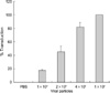
In order to determine the optimal transduction efficiency, varying numbers of viral particles were transduced. Transduction rate was defined by the percentage of cells staining blue with β-galactosidase activity. At viral particle numbers of 4×10⁸ and 1×10⁹ transduction rates of 82.4% and 100% were obtained, respectively (Fig. 1).
Determination of the effect of Ad5CMV-p53 infection on WB-2054-M5 cell growth
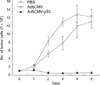
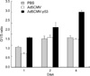
The hexosaminidase assay was used to determine the effect of Ad5CMV-p53 infection on WB-2054-M5 cell growth. Five days after the infection, growth of the tumor cells that were infected with Ad5CMV-p53 was significantly inhibited (Fig. 2). The cell growth assay reinforced findings in Fig. 2 that growth inhibition of Ad5CMV-p53-infected tumor cells was more significant, compared with the cells infected with only the viral vector Ad5CMV or only PBS (Fig. 3).
Effects of Ad5CMV-p53 infection on cell cycle and apoptosis in WB-2054-M5 cells
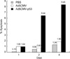
After transfection of p53 via the vector Ad5CMV-p53, WB-2054-M5 cells accumulate during the G1 phase as there was no progression into the S phase. By day 4 of incubation after transfection, the cells exposed to Ad5CMV-p53 had a significantly larger G1/S ratio compared to the cells infected with the viral vector alone or PBS (p<0.001) (Fig. 4). This is consistent with the belief that p53 inhibits the growth of WB-2054-M5 cells.
In addition to growth inhibition, tumor cells that were transfected with Ad5CMV-p53 had a markedly larger percentage of cells undergoing apoptosis than those transfected with Ad5CMV or PBS (p<0.001) (Fig. 5). To obtain an apoptotic index (apoptotic cells/total cells ×100), the apoptosis fluorescent staining assay was used to differentiate between viable and nonviable cells within a given population.
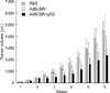
Effects of intratumoral injection of AdCMV-p53 on allograft colorectal tumor
Fig. 6 demonstrates significant suppression of allograft colorectal tumor after intratumoral injection of AdCMV-p53. The data was obtained from 54 rats, 18 from each of three treatment groups. PBS, Ad5CMV, or Ad5CMV-p53 were administered at 2-weeks following subcutaneous tumor cell injection. Tumor dimensions were recorded at a regular intervals of 3 or 4 days over a 6-week span. Tumor volume was obtained according to the ellipsoid formula. The volume of the tumors in the PBS, Ad5CMV, and Ad5CMV-p53 groups at week 4 and 6 was as follows: week 2: 1.66±0.43, 1.4±0.47, 0.75±0.26 cm3 (p<0.001), week 4: 4.41±0.88, 3.93±1.86, 2.33±0.51 cm3 (p<0.001). After adjustment for age, rat weight at operation and tumor volume at week 2, tumor growth of the Ad5CMV-p53 group was significantly suppressed (p<0.001). There was no significant difference between tumor volumes of the PBS and Ad5CMV groups (6-week vol. p=0.32). In the TUNEL assay, we found more apparent apoptotic cells in Ad5CMV-p53-treated tumors than in other groups (Fig. 7) 24 hr after the final treatment of 9 rats.
DISCUSSION
Colorectal cancer that metastasizes into liver remains one of the leading causes of cancer-related death in the United States. Research focus has been on the tumor suppressor gene p53 and its role in inhibiting tumor growth. p53 is mutated in about half of all human tumors and about 70% in colorectal cancers (7-9, 13). Mutant p53 has also been reported to decrease the expression of vascular endothelial growth factor (23). Mutation of p53 is considered to be an important tumor biology factor in colorectal cancer and other malignancies.
Various viruses and liposomes have been introduced as a means to deliver a specific gene. In particular, adenoviruses have been widely adopted as vectors for gene therapy in both animal and human trials. Drazan et al. (24) described the appeal of replication-defective adenovirus for use in hepatic gene therapy: 1) high purity concentrates of adenovirus vectors may allow for high multiplicity of infection in vivo, 2) the shuttle vector can insert DNA sequences into hepatic cells regardless of the organ's physiologic state, and 3) specific sero-types are highly hepatotropic and rapidly endocytosed by eukaryotic cells. Recombinant adenoviruses have various advantages over alternative delivery methods in that they usually do not insert their DNA into the genome of the host cell and that they are highly efficient modes of gene transfer.
Drazan et al. described the advantages of replication-defective adenovirus for hepatic gene therapy (24, 25). They emphasized the safety of adenovirus-mediated tumor-suppressor transduction into hepatocytes and over-expression of wildtype p53 transfected into quiescent and regenerating hepatocytes. Zhang et al. assessed the infectivity and cytotoxicity of Ad5CMV-p53 on human lung cancer cells and suggested that Ad5CMV-p53 at a dose that is effective in cancer cells is not detrimental to normal cells (26). The maximum tolerated dose of Ad5CMV-p53 in patients was the maximum attainable dose: 1×10¹¹ pfu. According to the data by Introgen Therapeutic, Inc., there was no treatment-related mortality or clinical signs of harm in rats and mice at dose of 1.43×10¹¹ pfu/kg. After direct intratumoral injection of recombinant adenovirus (total 2×10¹¹ vp/rat, 1 pfu=20 vp), there was no evidence of toxicity in the earlier mentioned in vivo experiments.
It was necessary to determine the ideal number of viral particles (vps) administered in order to get optimal transduction rates and to assess tumor cell growth in cells treated with Ad5CMV-p53. A transduction rate of 100% was obtained at the vp number of 1×10⁹. At this vp level, it was possible to assess growth of tumor cells using the hexosaminidase assay and the cell growth assay. Prior to performing the in vivo experiment, the ability of transfected wild-type p53 to suppress tumor cell growth can be assessed this way. WB-2054-M5 cells were more significantly inhibited by Ad5CMV-p53 than by PBS and Ad5CMV in both assays. In vitro cell proliferation analyses was done to evaluate the effect of adenovirus-mediated wild-type p53 expression. Theoretically, this analysis allows determination of whether the transfection of wild-type p53 suppresses tumor cells before performing the in vivo experiment. It is now well established that p53 induces growth arrest either at G1 or apoptosis (11). In the cell cycle assay, the G1/S ratio was markedly increased in the Ad5CMV-p53 group. This is consistent with the findings that p53 inhibits growth of WB-2054-M5 cells. Loss of viability, whether it be the result of a necrotic processs or apoptotic process, is often defined as loss of membrane integrity. Necrosis refers to the morphology usually associated with accidental cell death, while apoptosis is seen when cell death is programmed or physiologically regulated. Determination of whether a cell dies by apoptosis as opposed to necrosis can be best made by examining the distinct structural changes in the cell's chromatin, which occur prior to lysis of the membrane. The number of apoptotic cells in WB-2054-M5 cells after transfection of wild-type p53 increased and the apoptotic index markedly increased in the Ad5CMV-p53 group.
Many studies have been done to see the effect of p53 gene therapy on various tumors. Some researchers reported that growth of human head and neck cancer cells in nude mice was suppressed after the introduction of a wild-type p53 gene via a recombinant adenovirus (19, 27, 28). Ad5CMV-p53 has been shown to suppress growth of human lung cancer tumor in the orthotopic lung cancer model of nude mice (26). It has also been reported that Ad5-p53 suppresses the tumorigenic potential of human glioblastoma cells and inhibits cell growth in the rodent 9L glioma brain tumor (4, 13). Spitz et al. demonstrated that human colorectal tumor was significantly suppressed and apoptosis was induced in mice following direct intratumoral injection of adenovirus-mediated wildtype p53 gene (13). Ad5CMV-p53 inhibited tumor growth during and immediately after treatment. Harris et al. also showed that Ad5-p53 in vivo suppressed tumor growth and increased survival of nude mice bearing human colon cancer that expresses mutant p53 (29). The latter two studies were confined to xenograft subcutaneous tumors of colorectal cancer. In our experiments, a syngeneic rat model of allograft subcutaneous tumor was created and the tumor response after direct intratumoral injection of Ad5CMV-p53 for colorectal carcinoma was evaluated.
The positive results from the in vitro experiments encouraged the recombinant adenovirus-mediated p53 gene therapy in the syngeneic rat model for colorectal cancer. Tumor growth in the Ad5CMV-p53 group was significantly more suppressed compared to the other two groups. No difference in tumor growth was seen between the PBS and the Ad5CMV groups, indicating that toxicity to tumor cells was not mediated by the recombinant adenovirus alone. Harris et al. showed that intratumoral injection of recombinant adenovirus resulted in nonuniform transgene expression in subcutaneous tumors (29).
The TUNEL staining has been used for the in vivo analysis of apoptosis (13, 22, 30, 31). The TUNEL assay is based on direct, specific, in situ labelling of DNA breaks in the nuclei. It provides the in situ visualization of apoptosis at a single-cell level, while protecting tissue architectures. In our study, the TUNEL assay demonstrated more apoptotic cells in Ad5CMV-p53 treated tumors compared to the other two groups.
These results support a role for p53 gene therapy as an adjunct to current therapies available for cancers with mutations in this tumor suppressor gene. Ad5CMV-p53 demonstrated effective suppression of colorectal tumor in the allograft subcutaneous tumor model of rat. The important role of adenovirus-mediated wild type p53 gene transfer has been shown in several realms of tumor biology. Down-regulation of endothelial growth factor expression and thus inhibition of angiogenesis in human colon cancer can be achieved with wild type p53 gene transfer (23). This study employed advanced technology allowing for direct gene transfer into living animals and emphasized an opportunity to develop novel modalities of treatment for malignant tumors.


 XML Download