

 PDF
PDF ePub
ePub Citation
 Print
Print


INTRODUCTION
Cord blood (CB) has been successfully used as an important source of stem cell transplantation since 1988 (1-5). Although the limited cell dose of CB is a major obstacle for engraftment in the adult patients, CB can now be considered as an established stem cell source for transplantation in the pediatric setting. However, the delay for the engraftment procedure is still an important clinical problem for children, and the expansion of CB stem cells is of crucial importance in producing large numbers of hematopoietic progenitor cells and facilitating engraftment. The ex vivo expanded CB cells have been successfully engrafted into myeloablated animals, as well as adult human (6-9).
In most studies, the CD34+ cell selection was done before initiating cell culture (10), but the CD34+ cell selection itself is associated with a substantial loss of progenitor cells. Another practical issue is that because most CB units are stored in a cryopreserved state, the cryopreserved CD34+ cell selection is associated with increased cell losses as compared to unfrozen material (11, 12). Several studies have demonstrated that the hematopoietic potential of cryopreserved CB could be preserved, and the purification of CD34+ cells is not essential for the ex vivo expansion of CB (13, 14). Thus, if the absolute numbers of progenitor cells are increased after an ex vivo expansion of cryopreserved and unselected CB, as compared to fresh and selected CB, it would be much easier, more practical and cheaper for clinical applications.
Recent studies have also revealed that homing receptors and chemoattractants have an important association with the engraftment mechanism after stem cell transplantation (15, 16). If the numbers of progenitor cells as well as homing potential could be increased by the ex vivo expansion of cryopreserved and unselected CB, it would be beneficial for transplantation in adult patients, and it would also improve the engraftment speed.
We wanted to know whether the larger nucleated cell doses, as well as increment of stem cells that express homing receptors could be achieved by an ex vivo expansion of cryopreserved and unselected CB, and we also evaluated the cytokine combinations that are best for ex vivo expansion of CB.
MATERIALS AND METHODS
Cord blood collection and processing
Twelve CB samples were collected from an umbilical cord vein after full-term vaginal delivery, and they were placed into transfer bags containing acid citrate dextrose. An informed consent was obtained from all mothers. Red cells were depleted with 10% pentastarch (Jeil Pharm, Seoul, Korea) by a density gradient separation and the resultant leukocyte concentrates (LC) were cryopreserved, after the addition of a final concentration of 10% dimethyl-sulfoxide (Sigma, Sydney, Austrailia).
Thawing and ex vivo expansion
Frozen LC from CB were thawed in a water bath and washed by the method of Rubinstein et al. (17). The LC was seeded onto 6-well tissue culture plates at a concentration of 1×10⁵/mL in media supplemented with a combination of various cytokines, and the culturing was carried out without a medium exchange. After incubation for 2 weeks at 37℃ in 5% CO2 atmosphere, the cells were harvested and assayed.
Recombinant human cytokines
The following recombinant purified human cytokines were used in these studies: recombinant human (rh) stem cell factor (SCF; 20 ng/mL, Amgen, Thousand Oaks, CA, U.S.A.), rh thrombopoietin (TPO; 50 ng/mL, Amgen), rh flt3 ligand (FL; 50 ng/mL, Amgen), rh interleukin 6 (IL-6; 20 ng/mL, Amgen), and rh granulocyte colony-stimulating factor (G-CSF; 20 ng/mL, Amgen). The combination of cytokines for each 12 CB samples was as follows: SCF+TPO+FL (group 1), SCF+TPO+FL+IL-6 (group 2), and SCF+ TPO+FL+IL-6+G-CSF (group 3).
Clonogenic assays
Nucleated cells before cryopreservation and after 2 weeks of expansion were seeded onto methylcellulose medium (Stem Cell Technologies Inc., Vancouver, BC, Canada) at 4×10⁵/plate in duplicate and incubated for 2 weeks at 37℃ in humidified air and 5% CO2. The granulocyte-macrophage colonies of more than 50 cells were scored by the use of an inverted microscope.
Nucleated cell count and phenotype analysis
Total nucleated cell (TNC) counts and the phenotype analysis were performed before cryopreservation and after 2 weeks of expansion. TNC counts were performed using an automated cell analyser, Sysmex K-800 (Sysmex corporation, Kobe, Japan), and the mononuclear cells were isolated from the CB for a flow cytometric analysis. Dual-color flow cytometry of the CD34/CD38 cells, CD 34/CXCR4 cells, CD34/VLA4 cells, and the CD34/VLA5 cells (Becton Dickinson, San Jose, CA, U.S.A.) was performed using FACSort (Becton Dickinson). The cells were stained with the corresponding monoclonal antibodies for 45 min. After their incubation, the cells were washed three times in phosphate-buffered saline, fixed in 1% paraformaldehyde, and analysed by using Lysys II software (Becton Dickinson).
RESULTS
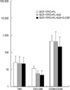
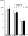
The absolute numbers of TNC, CFU-GM, CD34+CD38- cells, CD34+CXCR4+ cells, CD34+VLA4+ cells and CD34+VLA5+ cells after ex vivo expansion were compared to the absolute numbers before cryopreservation.
Effect of cytokines on the clonogenic potential after expansion of CB
The expansion folds for cell numbers for TNC were 23.2±13.8 (group 1), 22.3±7.8 (group 2), 26.3±4.9 (group 3); CFU-GM were 4.7±5.1 (group 1), 6.0±2.8 (group 2), 11.7±2.6 (group 3); CD34+CD38- cells were 214.0±251.9 (group 1), 464.1±566.1 (group 2), and 437.4±59.9 (group 3). No significant differences in extent of expansion of TNC, CFU-GM and CD34+CD38- cells were observed among the different cytokine combinations (Fig. 1).
Effect of cytokines on the homing potential after expansion of CB
The expansion folds for cell numbers were: CD34+CXCR4+ cells 4384.5±1664.7 (group 1), 6149.9±3552.0 (group 2), 7087.2±4669.3 (group3); CD34+VLA4+ cells 1444.3±1264.0 (group 1), 1832.6±1875.6 (group 2), 2074.9±1537.0 (group 3); CD34+VLA5+ cells 86.2±50.9 (group 1), 92.8±58.5 (group 2), 113.2±57.1 (group 3). No significant differences in extent of expansion of CD34+CXCR4+ cells, CD34+VLA4+ cells and CD34+VLA5+ cells were observed among the different cytokine combinations (Fig. 2).
DISCUSSION
There have been many studies regarding the growth factor combinations that could stimulate an optimum expansion of CB progenitor cells in stroma-free liquid culture. Among the various cytokines, FL, TPO, SCF and IL-6 seems to enhance the self-renewal and proliferative potential of primitive stem cells (18). Since we postulated that the additional presence of G-CSF might enhance the differentiation of expanded progenitor cells and increase TNC, which could facilitate the engraftment speed, we performed our present study with 5 cytokines including FL, TPO, SCF, IL-6 and G-CSF.
Almost all of the recent ex vivo expansion studies used freshly prepared or purified CD34+ cells from CB before cryopreservation (10, 18). Although Briddell et al. (10) demonstrated CD34+ cell selection is necessary for the optimal expansion of clonogenic cells, other studies have revealed that CD34+ cells and clonogenic cells could be expanded in unselected samples, as in contrast to the selected samples (13, 14). There may be some concerns regarding the detrimental effects of cryopreservation on the engraftment potential of expanded CB, however, DiGiusto et al. (8) and Rice et al. (19) have demonstrated that cryopreservation does not affect the engraftment potential of the frozen cells. Lazzari et al. (20) have also recently observed similar clonogenic efficiencies after ex vivo expansion of both fresh and cryopreserved CD34+ cells.
Since most of CB, that is used for clinical transplantation, is released from cryopreserved CB banks, it would be reasonable and practical to establish the protocol for the ex vivo expansion of CB from thawed and unselected state, and that is the reason why we performed our present study. We examined the increase of TNC, CFU-GM and CD34+CD38- cells by ex vivo expansion of cryopreserved and unselected CB. In contrast to TNC and CFU-GM, the CD34+CD38- cells, which are very immature progenitors and preserve their self-renewal capacity, they dramatically increased. There were no additive effects of using IL-6 and G-CSF on the expansion potential. We have demonstrated that the cell doses of immature progenitors could be increased by an ex vivo expansion of cryopreserved and unselected CB, and the combination of 3 cytokines (SCF, TPO, FL) is sufficient for this expansion.
There are several adhesion molecules necessarily involved in the mobilization and homing of CD34+ cells, such as CX CR4, VLA4 and VLA5. Since VLA4 is important for the early phase of lodgment of the CD34+ cells after transplantation, and the CD34+ cells that express high levels of VLA4 have more proliferative activities (16, 21), then the up-regulation of these adhesion molecules may be useful for improving engraftment in clinical transplantation. Recent studies have demonstrated that SCF, IL-6, IL-3 and granulocyte-monocyte colony-stimulating factor (GM-CSF) could induce the up-regulation of these molecules (22, 23). However, Ramirez et al. (24) have recently reported that although the expression of VLA4 and VLA5 was increased after ex vivo expansion, the adhesion of the progenitor cells to fibronectin was significantly decreased. A number of studies have also shown that the transplantation of ex vivo expanded progenitors has been associated with a delayed hematopoietic engraftment, that is, when the researchers transplanted fresh CD34+ cells or the equivalent numbers of expanded cells into irradiated NOD/SCID mice (16, 24, 25).
Yet, it would be very important to consider the absolute cell number, not the relative percentage of CD34+ cells expressing the marker when evaluating the clonogenic or homing potential after an ex vivo expansion. So, we evaluated the homing potential of expanded CB by comparing the absolute number of CD34+CXCR4+, CD34+VLA4+, CD34+VLA5+ cells after an ex vivo expansion and before cryopreservation. We have demonstrated a dramatic increment of the absolute cell numbers, and particularly for CD34+CXCR4+ and CD 34+VLA4+ cells, after an ex vivo expansion of the cryopreserved and unselected CB, and we also found that the combination of 3 cytokines (SCF, TPO, FL) is sufficient for a large increase of the stem cells expressing homing receptors.
Based on these data, we conclude that an ex vivo expansion of cryopreserved and unselected CB using the combinations of 3 cytokines (SCF, TPO, FL) could be sufficient for transplantation in adults. The number of stem cells expressing homing receptors could also be increased by an ex vivo expansion, and further in vivo studies regarding the engraftment potential after up-regulation of the homing receptors, as well as expansion of primitive stem cells will be required in the future.
 XML Download