

 PDF
PDF ePub
ePub Citation
 Print
Print


INTRODUCTION
DA-8159, a selective phosphodiesterase type 5 (PDE5) inhibitor developed by DongA Pharmaceutical Company (Kyunggi, Korea), is an oral agent for treating erectile dysfunction. DA-8159 induces penile erection dose-dependently in both anesthetized and conscious animals. It also induces smooth muscle relaxation and increases the endogenous cyclic guanosine monophosphate (cGMP) level in the rabbit corpus cavernosal smooth muscles (1). The data obtained from phase 1 clinical study showed DA-8159 is safe and well tolerated after a single oral dose in healthy males up to 300 mg without severe adverse effects (unpublished data). However, as with other PDE5 inhibitors, it may inhibit phosphodiesterase type 6 (PDE6) at a higher concentration. The inhibitory concentration of DA-8159 on the PDE6 receptor is 10 times higher than that of the PDE5 receptor.
PDE5 is present in human platelets and vascular smooth muscles. PDE5 inhibition causes a vascular dilatation by blocking cGMP hydrolysis in the vascular smooth muscle. PDE6 is present in retinal photoreceptor cells, and is essential for visual excitation, named phototransduction. The visual excitation begins with the absorption of a photon of light by the pigment rhodopsin. In this process, PDE6 hydrolyzes cGMP to guanosine monophosphate (GMP), resulting in a decrease in the intracellular cGMP levels. This light-dependent decrease in cGMP leads to hyperpolarization of the photoreceptors through the closure of cation channels. The inhibition of PDE6 increases the intracellular concentration of cGMP, which leads to opening of the sodium channels resulting in depolarization of the photoreceptor cells. The alteration of sodium channels causes exchange of Ca++, Na+ and Mg++ through the photoreceptor cells. As a result, ionic conductance generates an electrical response, which is transmitted to the visual cortex of the brain and produces a visual sensation. The visual excitation process can be recorded using electroretinography. If DA-8159 acts as a PDE6 inhibitor in retinal photoreceptor cells and inhibits the phototransduction process, an electrical alternation should be recorded in an electroretinogram (ERG).
Sildenafil citrate (Viagra®, Pfizer, Inc., New York, NY, U.S.A.) was initially developed as a drug to treat angina, but it was found to be highly specific to PDE5. Recently, it has been widely used to treat patients with erectile dysfunction. However, variable systemic and ocular side effects have been reported. The ocular side effects include visual halo (2), third nerve palsy (3), nonarteritic anterior ischemic optic neuritis (4, 5), etc. As observed with sildenafil, DA-8159 may cause such ocular side effects. Theoretically, PDE inhibitor may change the retinal physiology in two ways; an alteration of the phototransduction process by PDE6 inhibition at the photoreceptor cells, and an alteration in vascular flow by PDE5 inhibition at the vascular smooth muscle. We have previously been able to assess the alteration of phototransduction by ERG or the subjective visual symptoms, and the alteration of the blood flow by Doppler flowmetry (6-8).
The objectives of this animal experiment were to investigate the effects of DA-8159 on the ERGs, and to examine the histological change after DA-8159 administration in rabbits.
MATERIALS AND METHODS
Twenty male rabbits (1.5 to 2.0 kg of body weight, bw) were used for the electroretinography and blood concentration measurements. The rabbits were divided into four groups; the DA-8159 5 mg/kg, 15 mg/kg, and 30 mg/kg bw treated groups and a control group. The test drug, DA-8159, was dissolved in 5 mL of saline and fed through an L-tube. The control rabbits were given equal amount of saline. Each group consisted of five rabbits.
To evaluate the ERG changes after DA-8159 administration, electroretinography was performed prior to administration, one hour after, and five hours after the drug administration. To analyze the relationship between the blood concentrations of DA-8159 and the ERG changes, 5 mL of blood was drawn from the ear vein prior to and immediately after the ERG recording. The eyeball was enucleated immediately after electroretinography for the histological examination.
For electroretinography, the rabbits were kept in the dark for twenty minutes for adaptation. The pupil was dilated with an eyedrop of 2.5% phenylephrine hydrochloride. The animal was anesthetized with an intramuscular injection of ketamine hydrochloride (65 mg/kg bw) and xylazine hydrochloride (15 mg/kg bw) mixture. The recording electrodes were placed on both corneas. The ERG jet (Universo SA, Switzerland) was used as the recording electrode. The reference electrode was placed centrally on the shaven forehead. For the stability of the electrode, one end of a reference electrode was replaced by a skin needle, and was inserted into the forehead skin. The ground electrode was placed on the earlobe. Corneal dehydration was prevented by the frequent application of hydroxy-propyl-methylcellulose. The ERG was recorded using a UTAS-E 2000 system (LKC Technologies, Inc, Gaithersburg, Maryland, U.S.A.). The light stimuli were generated by a Ganzfeld dome stimulator (LKC Technologies). Full-field electroretinography was performed according to the Standard for Clinical electroretinography recommended by the International Society for Clinical Electrophysiology of Vision (ISCEV) (9). As recommended by the ISCEV, five basic responses were obtained from each rabbit. The significance of the ERG changes after drug administration was analyzed using a Bonferroni test. In brief, the time-dependent ERG changes according to the drug concentrations were compared with that of the control group. The relationship between the blood concentration of DA-8159 and the ERG changes were analyzed using linear regression analysis.
For histological examination, the eyeball was enucleated 1 or 5 hr after the drug administration. The eyeball was enucleated under general anesthesia with an intramuscular injection of ketamine hydrochloride and xylazine hydrochloride mixture. A total of twenty rabbits, two rabbits in each group, were used for the histological examination. After enucleation, the animal was sacrificed by ketamine overdose. Immediately after enucleation, the eyeball was immersed in a fixative solution of 2% glutaraldehyde in 0.1 M phosphate buffers. One eye from each rabbit was used for optical microscopic examinations, and the other was used for electron microscopic examinations. For the optical microscopic examination, the eyeball was placed in 20 mL of 10% formaldehyde for 24 hr. For electron microscopic examination, the eyeball was bisected and a tissue block was cut into a 2×3 mm size using an dissecting microscope. The block was placed in 2% glutaraldehyde in 0.1 M phosphate buffer for 90 min in the cold, and post-fixed in 1% osmium tetraoxide for 90 min. After fixation, the block was dehydrated serially with ethanol, and embedded in epon. A semi-thin section was stained with uranyl acetate and lead citrate. The ultrastructural study was performed by transmission electron microscopy (ISI-LEM 2000, Akashi, Japan).
RESULTS
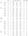
The five standard electroretinographic responses obtained from DA-8159 administrated and the saline ingested group are shown in Table 1. In the rod response, there was no remarkable ERG change at any dose or at any time after DA-8159 administration. In the maximal response, a statistically significant decrease in the b-wave amplitude (p<0.05) was noted at 5 hr after the administration of DA-8159 30 mg/kg, without a concurrent implicit time change. Otherwise no remarkable ERG changes were observed in the wave implicit time or amplitude at any dose or at any time after the test drug administration. The oscillatory potentials showed no significant change. In the cone response, there was no ERG change at any dose or at any time after the test drug administration. The 30-Hz flicker responses showed a statistically significant prolongation in the implicit time (p<0.05) after 5 hr of DA-8159 15 mg/kg or 30 mg/kg bw administration. The changes were associated with a concurrent decrease in the amplitude, even though this was statistically not significant (p>0.05).
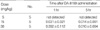
The dose-dependent blood concentrations of DA-8159 as a function of time are shown in Table 2. No test drug was detected in the blood after 1 or 5 hr at dose of DA-8159 5 mg/kg bw. At doses of 15 mg/kg and 30 mg/kg bw, there was a dose-dependent increase of the blood concentration after 1 hr of drug administration. However, no difference in the blood concentration was found after 5 hr. There was no correlation between the blood concentration and the ERG change after 1 or 5 hr of test drug administration.
No abnormal light and electron microscopic findings were observed in the DA-8159 treated groups. No abnormal findings were observed either in the sensory retina, the photoreceptor rod and cone cells, the retinal pigment epithelial cells, the Bruch's membrane, or the choroidal vessels. An electron microscopic examination showed the vascular endothelial cells of the choroid exhibit well-preserved terminal bars connecting the adjacent endothelial cells.
DISCUSSION
ERG has established itself for many decades as a routine diagnostic method in clinical ophthalmology. It is a summation of the electrical responses generated by the neural and non-neuronal cells within the retina. However, it is necessary to standardize the recording and interpreting protocols in order to make it possible to compare the results elicited in different institutions. In 1989 the ISCEV provided standard ERG test procedures and five basic responses (9). The five basic ERG responses are the rod response, the maximal rod and cone response, the oscillatory potentials, the cone response, and the 30-Hz flicker response. However, the standardization is suitable for humans, but not for rabbits. The anatomical and functional similarities of the rabbit retina have made it a simulated model for an ophthalmic study, but the retinal anatomy of the rabbit is slightly different from that of humans. There are three types of cone cells in the human retina, but only two, green cones and blue cones (10, 11), are present in the rabbit retina. In addition, there is no standardized data for the rabbit ERG, which makes it unadvisable to use human ERG data in rabbits. Compared to the baseline values, a statistically significant difference was observed between each group. Therefore, it is advisable to compare the time-dependent ERG change in the given group with that of the control group. In this study, the statistical significance of the time-dependent ERG change was analyzed by comparing the data from the test drug administrated group with that of the control group.
Luu et al. (12) studied the effect of sildenafil on the full-field ERG, color vision and the subjective visual symptoms on healthy volunteers. Two out of fourteen subjects who received 200 mg of sildenafil complained bluish vision. Those who showed a depression in the ERG cone function made more errors in the color vision test. In addition there was a correlation between the ERG changes and the occurrence of the subjective visual symptoms. They made a conclusion that 200 mg sildenafil caused rather mild acute alterations in the cone and rod function. Lee et al. (13) reported an alteration of ERG after a single dose of 100 mg sildenafil ingestion. Similar ERG changes were found in animal experiments. There was statistically significant prolongation of the implicit time in the 30-Hz flicker response at doses of 15 mg/kg bw and 30 mg/kg bw DA-8159 with a concurrent decrease in amplitude, although this was statistically insignificant. Similar ERG changes were observed in the cone response at 30 mg/kg bw. Such ERG changes were not observed at 5 mg/kg bw. Because the 30-Hz flicker response or cone response represents the photoreceptor cone function, its alteration may cause color vision changes. These ERG changes are useful to explain the pathogenesis of color vision alterations after PDE inhibitor administration.
The mechanism of the ERG changes after PDE inhibitor administration is still unclear. Theoretically a higher concentration of a PDE inhibitor may cause an alteration in the phototransduction process, resulting in the ERG changes. Behn et al. (14) investigated the effect of sildenafil on the retina in knockout mice, which were heterozygous for a mutation causing an absence of the γ subunit of rod PDE6. They found that sildenafil significantly decreased the a-, and b-wave amplitudes in the heterozygous PDE6γ subunit lacking mice. Further decrease in both the a-, and b-wave amplitude were observed with increasing sildenafil doses. The ERG decrease was more pronounced in the heterozygous PDE6γ subunit lacking mice than in the wild mice containing normal PDE6. This result shows that the sildenafil-induced ERG change is not related to the PDE6 inhibition caused by sildenafil. However, they asserted that the heterozygous PDE6γ subunit knockout mutation probably leads to a decrease in the amount of functional PDE6, creating an enhanced susceptibility to the inhibitory effects of sildenafil. In contrast, Vobig et al. (15) reported significant reductions in the a-, and b-wave amplitudes 1 hr after administering sildenafil, and these effects recovered to normal levels after 6 hr. The amplitude reduction correlated well with the slidenafil plasma concentration, which showed a peak 1 hr after administering the drug. Moreover, Behn et al. (14) reported that sildenafil has a significant dose-dependent inhibitory action on the retinal function. The retinal inhibitory effect occurred with as little as twice the maximum equivalent dose recommended for humans. These results indicate a close correlation between the sildenafil blood concentration and the ERG change. However, this study found no significant ERG change at 5 mg/kg bw DA-8159, which was 3.5 times higher-dose recommended for a 70 kg person.
In this animal study, no correlation between the DA-8159 blood concentration and ERG changes could be found. The mean blood concentration at 1 hr after DA-8159 15 mg/kg bw administration was as high as 2.2 times than that observed 5 hr after (0.031 g/mL vs. 0.014 µg/mL, p<0.05), but the ERG amplitude changes were the opposite. There was a statistically significant ERG cone response change 5 hr after administering the test drug, not after 1 hr. As provided by manufacturer, the Tmax of blood DA-1859 is approximately 60 min after ingestion. If the ERG changes are due to the PDE6 inhibition effect of DA-8159, the changes will peak around 1 hr after administration, not 5 hr after. This suggests no significant correlation between the DA-8159 blood concentration and the ERG changes. Schneider et al. (16) reported a change in the ERG response related to the dose of the PDE inhibitor in cats. The phosphodiesterase inhibitor increased the rod b-wave amplitude at low concentrations, but diminished the rod b-wave amplitude at high concentrations. In view of the results, it is possible that there is a correlation between blood concentration of PDE inhibitor and the ERG change at relatively high doses, positively or negatively. However, there is some uncertainty in the correlation between the blood DA1859 concentration and the ERG change.
In conclusion, there were no significant ERG changes after administering 5 mg/kg bw DA-8159. However, at 15 mg or 30 mg/kg bw, a statistically significant prolongation of the 30-Hz flicker implicit time was observed 5 hr after the test drug administration. Furthermore, there was a b-wave amplitude decrease in the cone response at 15 mg or 30 mg/kg bw, although this was statistically insignificant. Otherwise, no remarkable ERG changes were observed in the rod, the maximal, and the oscillatory potentials. There was no correlation between the blood DA-8159 concentration and the ERG change. In light and electron microscopic examinations, there were no histological changes after DA-8159 administration at any dose or at any time. These data suggest DA-8159 has a minimal effect on ERG changes in rabbits, but further evaluation of the effects of DA-8159 on visual functions in human must be followed.
 XML Download