

 PDF
PDF ePub
ePub Citation
 Print
Print


INTRODUCTION
Kawasaki disease (KD) is an acute multisystem vasculitis that afflicts mostly young children. Since the first report of KD in 1967 (1), many attempts to determine the etiology of the disease have failed. However, the clinical and epidemiologic studies have proposed that KD is closely related to an infectious disease (2, 3). The acute onset of a self-limited course, the prevalent population (rare in <6 months of age and >5 yr of age), the existence of clusters or epidemics with a wave-like spread, all suggest that KD is related to infectious agents, particularly of viral origin. On the other hand, the elevated levels of the inflammatory indices including the white blood cell (WBC) and neutrophil count and the C-reactive protein (CRP) suggest that bacterial agents including superantigens are involved in KD (4). We previously found that the IgM and IgA levels increased at 1 week and 2 weeks after intravenous immunoglobulin (IVIG) treatment with a statistical significance (5). This finding also provides evidence showing that KD is an infection-related disease.
Many therapeutic modalities have been attempted in order to prevent the coronary artery lesions (CAL) as the major complication of KD. IVIG therapy is known to decrease the numbers of CAL, and is now accepted as standard treatment for KD (6, 7). Therefore, a study on the natural course in KD is unethical and impossible.
The total duration of fever in KD in the era before IVIG therapy was reported to be approximately 1-2 weeks (mean 10 days) regardless of treatments with aspirin or steroids (1, 8).
We evaluated the inflammatory indices including the WBC count and CRP according to the onset of fever in children with KD, and postulated that inflammatory processes in KD reach a peak at the sixth day of fever.
MATERIALS AND METHODS
The subjects of this study were 152 children who had been diagnosed with KD (82 boys and 70 girls) between July 1999 and December 2002 at The Catholic University of Korea, Daejeon St. Mary's Hospital. The diagnostic criteria were based on the Diagnostic Guidelines of Kawasaki Disease presented by the Japan Kawasaki Disease Research Committee (9). The first day of fever was considered the first day of illness. The mean age of the patients was 2.4±1.4 yr (2 months-9.4 yr). One hundred nineteen children met the criteria for KD and 33 cases of an incomplete KD were included. Incomplete KD patient was defined as those who do not fulfil the recommended criteria at presentation regardless of echocardiographic findings. All patients with incomplete presentation who did not show elevated CRP (<2.0 mg/dL), and WBC and neutrophil (<10,000/µL and <5,000/µL, respectively) levels with repeated examinations in the earlier days of illness or those who did not show an increased platelet count 7 days after IVIG treatment were excluded (4 cases). Children were treated with IVIG (I.V.-Globulin S, Green Cross, Korea: 5% liquid preparation containing only maltose, IgG:maltose=1:2) at a dose of 2 g/kg over 12 hr and a dose of aspirin (30-40 mg/kg) during the febrile period. After obtaining parental consent, the serial examinations were performed three times during admission: before IVIG administration, and 24 hr and 7days after IVIG administration. CAL were defined and classified as follows: ectasia was defined when coronary arterial dilatation with the diameter ≤4 mm was seen or when the diameter was less than 1.5 times than that of adjacent artery diameter; aneurysm, when dilatation >4 mm ectasia with or without, multiple, pyramidal/fusiform aneurysm was present. Twenty-six of the 152 children had CAL (21 in ectasias and 5 in aneurysms). Thirteen children showed a resistance to IVIG therapy (a fever for more than 48 hr after initiating the IVIG infusion). These children were not excluded from the study group. The demographic and laboratory data were tabulated. The Ethics Committee on Clinical Research, the Catholic University of Korea, approved this study.
Statistical analyses were done using SPSS 10.0 for Windows. The means of all continuous variables were compared using one way ANOVA, Fisher's extact test and chi-square test. Continuous variables are reported as the mean±Standard deviation. p≤0.05 was considered statistically significant.
RESULTS
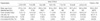
The demographic and clinical characteristics of the patients are summarized in Table 1. There were no significant differences in terms of age (mean 28.4±16.2 months) and sex distribution (male to female ratio, 82:70) among the groups. The mean total duration of the fever was 7.3±1.9 days. The mean incidence of CAL evaluated within 2-3 weeks of the onset of fever was 17.1%. There was a trend for the incidence of CAL to be higher in the sixth day (40%) and the ≥ninth day groups (42.9%) than the other groups. No significant differences were observed among the groups in the cases of IVIG retreatment. Twenty-two percent of cases had fever with less than 4 of the diagnostic criteria for KD at presentation (incomplete KD). There was a trend for the incidence of incomplete KD to be higher in the ≤third day (35%), the eighth day (33%) and the ≥ninth day groups (43%).
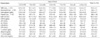
The laboratory findings obtained before IVIG treatment are summarized in Table 2. WBC and neutrophil counts, CRP and creatine phosphokinase (CPK) values were the highest in the sixth day group, and the last two indices showed a bell-shaped distribution pattern based on the peak sixth day values. In contrast, albumin, and HDL-cholesterol values were the lowest in the sixth day group, and they showed a pattern of U-shaped distribution. The platelet count and total cholesterol value showed a trend for increase with subsequent days of fever. The hemoglobin, ESR and LDH values did not change significantly. The AST and ALT values were the highest in the ≤third day group, but were not statistically significant.
DISCUSSION
The inflammatory processes of an infection progress to a peak stage, then regress to a convalescence by host immune response. The total duration of fever in most uncomplicated viral infections is approximately 1 week, including measles (5 days) and Epstein-Barr virus infection (6 days). On the other hand, the duration of fever in an untreated bacterial infection differs according to the causative agent. Scarlet fever is known to last 5-7 days. However causative disease caused by intracellular organisms such as typhoid fever lasts much longer (>2 weeks).
Early studies revealed that the total duration of KD without IVIG therapy was 1-2 weeks (mean 10 days) (1, 8). Therefore, it is postulated that the peak inflammatory process in KD is at the fifth to sixth day after the onset of fever. The results in this study shows that the levels of inflammatory indices reach a peak or nadir on the sixth day of the fever, which agrees with the above postulation. WBC and neutrophil counts, ESR and CRP levels are commonly used as indices of severity of inflammation. CRP increases rapidly within 24 hr in various conditions, like bacterial infections, trauma, tissue necrosis, and malignant neoplasm. It declines rapidly with a resolution of the pathologic conditions (10). ESR is also another acute reactant of inflammation. The decreased albumin or HDL-cholesterol values have been reported in inflammatory diseases including KD (11, 12). Although, this study could not show difference of statistical significance in any of the indices between groups, the distribution patterns of these indices strongly support our postulation. AST and ALT values appeared to be higher in the early days of the natural course in KD. The cases with elevated AST and ALT above two folds the normal values were also higher in the early days of the fever (≤4 day, 20 of 50 cases) than in the later days (≥7 days, 5 of 38 cases). A decreased total cholesterol level in the earlier days tends to increase with days of the fever. The platelet count is well known to increase in the convalescent stage in KD. The platelet counts also tended to increase by days of fever in the acute stage.
Recently, two studies reported the relationship between the initial day of the IVIG treatment and the clinical or laboratory outcomes in KD (13, 14). Although the two studies are similar in study design, the results of CAL frequency and laboratory findings were not identical. Nomura et al. (13) reported that the group receiving IVIG treatment prior to the fifth day of illness showed a higher CAL at 1 month, and no differences in the laboratory findings except for ALT and AST when compared to those given IVIG after the fifth day. In contrast, Tse et al. (14) reported a case-control study showing that early IVIG treatment at day 5 or earlier showed a lower incidence of CAL at 1 yr, and some differences in the laboratory findings including higher hemoglobin, ALT, and albumin values and lower platelet count. If our postulation is correct, different results of laboratory study between the two groups might result from differences in the number of patients in each different fever day.
Earlier studies have reported that the risk of CAL in KD is associated with some demographic or laboratory factors such as the prolonged fever duration, or an increased CRP (15-17). IVIG is quite effective for improving the clinical symptoms as well as the laboratory findings including CRP, and for preventing coronary complications (6, 7). We previously repored that a high dose IVIG treatment (2 g/kg) lowered the values of various proteins including albumin and lipoproteins, and also values of the inflammatory indices within 24 hr except ESR (5, 18). In addition, we also found that IVIG-resistant patients showed a sustained high values of CRP and WBC on 24 hr after IVIG with a higher risk of CAL (19, 20). If the risk of CAL is associated with the intensity of the inflammation reflected by laboratory indices, early treatment prior to the peak stage of inflammation can help prevent CAL in KD.
There are some limitations in interpreting our results. Because the data had a large number of variables (seven variables) and an uneven distribution of the number of variables, our postulation could not reach statistical significance. A prospective study with a large number of patients would resolve this. A diagnosis of KD in patients before 5 days illness could not fulfil the diagnostic criteria and those with incomplete KD were included in this study. All patients with incomplete KD were selected on the basis of the laboratory findings as described earlier as well as the clinical criteria. However, there was no significant difference in laboratory values between the incomplete KD group and typical KD group (data not shown).
In conclusion, it is very important to determine when the inflammatory processes of KD reach a peak as a self-limiting disease. As for the coronary complications in KD, it is believed that the more severe inflammatory processes, which are reflected by prolonged fever duration or higher values of the inflammatory indices, have a higher risk. Therefore a higher single-dose IVIG treatment before the peak stage of KD may help reduce the intensity of inflammation and the frequency of CAL. Repeated examination of the inflammatory indices can help decide the timing for appropriate IVIG treatment for children with an incomplete presentation in the earlier days, and for evaluating the effects of IVIG treatment.
 XML Download