

 PDF
PDF ePub
ePub Citation
 Print
Print


INTRODUCTION
Epithelioid hemangioendothelioma is a rare vascular tumor that usually develops in the lung, liver, bone, and soft tissue, and has a prognosis varying from benign to malignant. Hepatic epithelioid hemangioendothelioma usually occurs in adult women and presents as multiple masses, and its confirmatory diagnosis is made by biopsy (1). Several cases have been reported in Korean literature, too (2).
On the other hand, hypertrophic osteoarthropathy is a syndrome characterized by periosteal new bone formation, clubbing and joint effusion. It develops as a primary disease or secondary to chronic pulmonary disease, cyanotic heart disease, inflammatory bowel disease, or chronic liver disease (3). We recently experienced a rare case of hypertrophic osteoarthropathy which developed in association with epithelioid hemangioendothelioma.
CASE REPORT
A 42-yr-old man visited our rheumatology clinic because of bilateral ankle arthritis in May 2002. He was diagnosed as having chronic hepatitis B in 1992, and multiple liver masses were found incidentally in July 1996. A liver biopsy performed at the time revealed prominent epithelioid cells containing the factor VIII-related antigen. These findings were consistent with epithelioid hemangioendothelioma in the liver. He received conservative management. He had no musculoskeletal complaints or abnormal physical findings at that time.
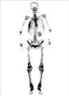
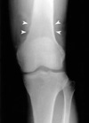
Pleural effusion and several lung masses developed in the right lung in July 2000. Biopsy findings of the pleural mass showed dysplastic epithelioid hemangioendothelioma with pleomorphisms, mitoses, and necrosis. It was speculated that the liver masses had metastasized to the right lung and the pleura. In April 2002, he began to feel pain and swelling in both ankle joints, which then extended to both wrists and knees. He visited our clinic because of this increasing pain and swelling of affected joints. Physical examination revealed swelling and tenderness of both ankle and wrist joints and mild tenderness of the right knee joint. Pitting edema was found in both lower legs, and prominent clubbing was found in all fingers of both hands. Laboratory data showed hemoglobin 13.6 g/dL, white blood cell count 5,700/µL, platelet count 182,000/µL, erythrocyte sedimentation rate 35 mm/hr (reference range, 0-10 mm/hr), alkaline phosphatase 180 IU/L (reference range, 30-115 IU/L), AST/ALT 35/38 IU/L (reference range, 0-40 IU/L, 0-40 IU/L, respectively), negative rheumatoid factor, and negative antinuclear antibody. Bone scan showed diffuse cortical uptake in the distal parts of both femurs and tibias (Fig. 1), and plain radiography showed subperiosteal new bone formation in the same areas (Fig. 2). Immunohistochemistry staining of the tumor tissue showed the presence of vascular endothelial growth factor (VEGF) (Fig. 3). A diagnosis of hypertrophic osteoarthropathy was made, and naproxen was prescribed, which relieved the arthritis and swelling in both lower legs. However, he was readmitted because of radiation-resistant metastatic lung lesions in April 2002 and was discharged hopelessly.
DISCUSSION
A diagnosis of epithelioid hemangioendothelioma was made on our patient, based on the typical biopsy findings of the dysplastic proliferation of factor VIII-related antigen positive cells. Epithelioid hemangioendothelioma was first reported by Dail and Liebow in 1975 as an intravascular sclerosing bronchoalveolar tumor of the lung. In the following studies, features of endothelial cells were observed in round or cuboid tumor cells on electron microscopy (Weibel-Palade body) and on immunohistochemistry (factor VIII-related antigen, von Willebrand factor). These findings suggest that epithelioid hemangioendothelioma originate from endothelial progenitor cells (4).
The case described in this report was diagnosed as hypertrophic osteoarthropathy based on findings of clubbed fingers, arthritis, and periosteal reactions, which were confirmed by typical radiographic and bone scan findings. His arthralgia responded very well to nonsteroidal anti-inflammatory drugs. The pathologic findings of hypertrophic osteoarthropathy are known to show vascular hyperplasia and activated endothelial cells in clubbed fingers, and vascular hyperplasia and periosteal proliferation in affected bones. In cases of cyanotic heart disease, the activation of cytokine-producing endothelial cells by macrothrombocytes is found, which might be a mechanism of hypertrophic osteoathropathy (5). Plasma levels of the von Willebrand factor antigen, which is a marker of endothelial cells, is known to be elevated in hypertrophic osteoarthropathy (6). These findings suggest that the activation of endothelial cells and platelets plays an important role in the pathogenesis of hypertrophic osteoarthropathy. Recently, the plasma and serum levels of VEGF, a hypoxia-inducible cytokine secreted by various cells including platelets, were found to be increased in patients with hypertrophic osteoarthropathy. The ability of VEGF to promote angiogenesis and the differentiation of osteoblasts (3, 8) suggests a role of VEGF in the development of hypertrophic osteoarthropathy.
The endothelial origin of epithelioid hemangioendothelioma suggests a link between the tumor and hypertrophic osteoarthropathy. Recently several reports have suggested an association between epithelioid hemangioendothelioma and VEGF. VEGF and VEGF receptors were found in a child case of epithelioid hemangioendothelioma (9), and VEGF blood levels were found to be reduced after tumor treatment with interferon-α (10). These findings suggest that VEGF play an important role in the pathogenesis of epithelioid hemangioendothelioma and hypertrophic osteoarthropathy in our patient. Consistent with this hypothesis, we observed the presence of VEGF in the tumor tissue of our patient (Fig. 3).
To the best of our knowledge, two case reports of epithelioid hemagioendothelioma complicated by hypertrophic osteoarthropathy have been reported. In one report, hypertropic osteoarthropathy was found in a 17-yr-old boy, concurrently with hepatic and pulmonary epithelioid hemangioendothelioma (11). In the other, hypertrophic osteoarthropathy was diagnosed in a 24-yr-old woman concurrently with pulmonary epithelioid hemangioendothelioma (12). Our case is the first Korean case of hypertrophic osteoarthropathy developing in the presence of epithelioid hemangioendothelioma. This case differs from previous reports in that the tumor was first found in the liver and hypertrophic osteoarthropathy developed after pulmonary metastasis, thus emphasizing the implication of a tumor in the lung in the pathogenesis of hypertrophic osteoarthropathy. Furthermore, our case suggests that hemangioendothelioma, albeit rare, should be included in the differential diagnosis of hypertrophic osteoarthropathy.

 XML Download