

 PDF
PDF ePub
ePub Citation
 Print
Print


INTRODUCTION
Bronchogenic cysts are derived from the embryologic branchial cleft and are mainly of pulmonary origin. They are rarely located in an extrathoracic site, such as subdiaphragmatic retroperitoneal area (1-17). Only a few cases of intraperitoneal area (18-26) have been documented (Table 1). To the best of our knowledge, only 22 retroperitoneal cases have been reported in the world literature by the year of 2001, 17 of which are English language reports (17). Cases arising from an intraperitoneal position are more unusual. Only 8 cases have been reported by the year of 2001. We report upon the first isolated intraperitoneal bronchogenic cyst in a 48-yr-old woman, which was presented as a gallbladder mass in Korea.
CASE REPORT
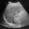
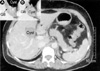
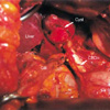
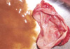
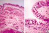
A 48-yr-old female was admitted to our hospital with one-year history of dyspepsia after meals and intermittent epigastric pain. A physical examination demonstrated no palpable mass in the abdominal region. White blood cell (WBC) count was at 5.2×10⁹/L, and hemoglobin was at 11.9 g/dL. Blood chemistry results were normal and preoperative serum alpha-feto protein (AFP) was also within normal range (0.77 U/mL, normal 0-5 U/mL). Ultrasound sonography showed a cystic mass adjacent to the gallbladder (Fig. 1). Abdominal CT showed a well defined and circumscribed, cystic mass 3×2.5 cm in size at the inferomedial aspect of the gallbladder (Fig. 2). Radiological findings suggested a gallbladder tumor, a teratoma, bronchopulmonary sequestration, a complicated cyst or carcinoma, but the findings were insufficient for an accurate diagnosis to be made. Therefore a presumptive diagnosis of a gallbladder tumor was made. The lesion was explored because CT did not show a definite demarcation between the mass and the neighboring structures, nor did it confirm its isolation in the gallbladder area; moreover, the possibility of malignancy could not be ruled out. At laparotomy, a 3 cm-sized cystic mass was discovered adherent to the gallbladder (Fig. 3). The cyst was dissected from the liver bed, and the entire cyst and gallbladder were excised consequently. There was no connection between cyst and gallbladder. The gross appearance of the resected specimen seemed to be a benign cyst. On opening the specimen revealed one large cystic cavity, which contained thick brownish mucoid fluid (Fig. 4). Microscopically, the cyst is lined by a layer of pseudostratified ciliated columnar epithelial cells occasionally interspersed with goblet cells (Fig. 5). Thus, the cyst was histologically diagnosed as a bronchogenic cyst. The postoperative course was uneventful; the patient was discharged at 10th day postoperatively, and had remained asymptomatic through biweekly follow-ups for two months.
DISCUSSION
Bronchogenic cysts are congenital abnormalities arising from the ventral foregut during the third to seventh week of fetal development. They are almost always lined, at least partially, by ciliated cuboidal to pseudostratified columnar epithelium and are often filled with mucus. Bronchial components such as cartilage, smooth muscle, elastic fibers, fibrous tissue and seromucinous glands may all be presented in the cyst wall (27). A retroperitoneal location is rarely reported. Although the exact mechanism is unknown, Sumiyoshi et al. (2) proposed the following theory. During early embryonic life, the thoracic and abdominal cavities are linked via the pericardio-peritoneal canal. When the canal is later divided by the fusion of the pleuroperitoneal membranes (the future diaphragm), a portion of the tracheobronchial tree may be pinched off and migrate, resulting in a retroperitoneal bronchogenic cyst (2). However, subdiaphragmatic bronchogenic cysts, especially in the intraperitoneal region, are extremely rare. Only 8 cases have been reported in the world literature, and all had their locations adjacent to the stomach. Our case had an unique gallbladder location. To our knowledge, no intraperitoneal cyst arising near the gallbladder had been reported in either the Korean or the English literatures. Of these retroperitoneal bronchogenic cysts, nine cases occurred in males and eight in females. The age of the patients varied because the cases of smaller cysts were asymptomatic and the masses were incidentally discovered. In the cases of larger cysts, the patients complained of various types of pains in the suspected region. The size of the cyst showed a increasing tendency with ages of the patients. Table 1 summarizes the eight cases of isolated intraperitoneal bronchogenic cysts that have been reported by the year of 2001. Interestingly, eight cases were located adjacent to the stomach. All of the eight cases were considered to arise in the left side of stomach. Preoperative clinical diagnosis included the followings; benign tumor (18), leiomyoma or lipoma (19), a intestinal obstruction (20), and a dermoid cyst (24). In our case, which located beside the gallbladder, apart from stomach, and no connection to stomach and gallbladder wall.
Since there are no common symptoms and specific changes in laboratory findings, CT scan has an important role in making the diagnosis. CT scan is useful for evaluating cyst contents so it allows a further differential diagnosis of retroperitoneal cystic lymphangioma, hematoma, abscess, etc. (28). Bronchogenic cysts usually show high CT values ranging from 30 to 100 HU since the cysts are filled with protein-rich fluid (12).
For histological diagnosis, they should be differentiated from bronchopulmonary sequestration and cystic teratoma. Bronchopulmonary sequestration can be diagnosed by the fact that it cintains has lung parenchyme and pleural tissue. Cystic teratoma has endoderm-origin bronchial tissue and other structures from mesoderm and ectoderm. Among the cysts of foregut origin, those containing cartilage or seromucinous respiratory glands are designated as bronchogenic cysts; those containing two well-developed layers of smooth muscle without cartilage are designated as esophageal cysts; and those with none of these distinguishing features are classified as foregut cysts (9). In contrast, the cysts of urogenital origin may rarely have pseudostratified ciliated epithelium, and submucosal seromucinous glands (4, 12). In our case, a teratoma was excluded by the absence of tissue, representing the three different germinal layers. In addition, bronchopulmonary sequestration can be diagnosed by the fact that it possesses lung parenchyma, pleural investment, and bronchial elements which were absent in our case.
Preferred treatment of intraperitoneal bronchogenic cyst is surgical removal. Although most are asymptomatic, excision is recommended to establish the diagnosis, alleviate symptoms, and to prevent complications, such as infections and the remote, but documented risk of malignant transformation (16).
Although the occurrence of bronchogenic cyst is rare, it should be considered in the differential diagnosis of an intraabdominal mass, particularly in the case of a cystic tumor in the region adjacent to the gallbladder.
 XML Download