

 PDF
PDF ePub
ePub Citation
 Print
Print


INTRODUCTION
Endocarditis related to pacemaker lead infection is a rare, but serious condition in permanent venous tracing. Staphylococci are involved in the majority of these infections (1-4) and Achromobacter xylosoxidans is very rare cause of organism. Although some studies reported that medical extraction of even large vegetation appeared to be safe (5), usually the infected endocardial system must be entirely removed and appropriate antibiotic therapy pursued for 6 weeks (1, 6).
CASE REPORT
A 35-yr-old patient visited our hospital due to a unremitting high fever and chills for 10 days. He had undergone a patch closure of the ventricular septal defect (VSD) 18 yr before. One year later, a VVI pacemaker was implanted via the right subclavian vein because of complete heart block. Nine years after that, a new VVI pacemaker with another right ventricular electrode was inserted controlaterally and the old pacing lead was abandoned. Several months before admission, he had the scaling and root planning at local dental clinic. He did not have any intravenous treatment history.
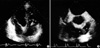
On admission, vital signs were blood pressure 120/75 mmHg, body temperature 39℃, and pulse rate 75/min. Physical examination revealed systolic ejection murmur in the left second intercostal area. The results of a complete blood count were normocytic, normochromic anemia, and leukocytosis (15,400/µL). An elevated erythrocyte sedimentation rate (ESR) (135 mm/hr) and positive C-reactive protein (CRP) (13.5 mg/dL) were demonstrated. Chest radiography revealed a mild cardiomegaly without active lesions in the lung parenchyma. Blood cultures were positive for Achromobacter xylosoxidans (colorless growth on MacConkey agar, oxidase positive, indole negative, saccharolytic, motile, rod-shaped nonfermenters analyzed by Vitec) 5 times. Susceptibility study showed that the strain was susceptible to ceftazidime, piperacillin, cefoperazone, imipenem, trimethoprim-sulfamethoxazole, and was resistant to aminoglycosides. Trans-thoracic echocardiogram (TTE) (Fig. 1A) and trans-esophageal echocardiogram (TEE) (Fig. 1B) identified the pacemaker lead in the right ventricle (RV) attaching hyperechoic materials and also a fluttering round hyperechoic mass with a stalk in the RV outflow tract (RVOT). No shunt flow was detected through the previous patch closure site in TTE and TEE. The function of pacemaker was normal on pacemaker analysis and 24-hr Holter monitoring.
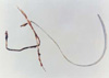
Despite sensitive antibiotics of ceftazidime and piperacillin for 3 weeks, patient's status was not stabilized. Therefore, the patient underwent a cardiac surgery through median sternotomy under extracorporeal circulation. Intraoperatively, yellow-brown friable materials attached to the older electrode (Fig. 2) and the septal wall of RVOT were confirmed. Removal of infected wires, infected Dacron patch, and vegetations in the RVOT, and curretage of infected RV wall were performed. Cultures for Achromobacter xylosoxidans were positive in the pus attached to the pacemaker lead. With sensitive antibiotics treatment, the patient was doing well 6 weeks postoperatively without any signs for infection. Epicardial pacing was maintained. There were no adverse changes in the normalized CRP and ESR. Thus, a transvenous permanent pacemaker implantation was performed after confirming the patency of the left subclavian vein with venography.
A follow-up echocardiogram 1 yr later demonstrated no detectable vegetations. The patients has been doing well without recurrence of infection for 2 yr.
DISCUSSION
Infection of the pacemaker pouch and lead may occur in 1% to 7% of patients with a permanent pacemaker (3). The microorganism most responsible for a late pacemaker lead infection is Staphylococcus epidermidis (4). Achromobacter xylosoxidans is a nonfermentative Gram-negative bacilli. This organism is opportunistic and usually affects severely immunocompromised patients such as those with neutropenia and those with a malignant or cardiovascular disease (7, 8). To the best of our knowledge, however, the present case is unique in that there has been no report such as ours. A case report of aortic valve and VSD Dacron patch infective endocarditis due to Achromobacter xylosoxidans 15 yr after a VSD patch closure appeared on Medline search (9). He did not have any definite cause of pacemaker lead endocarditis. Several months before admission, he had the scaling and root planning at local dental clinic.
The diagnosis is difficult by using conventional imaging methods such as transthoracic echocardiogram. TEE can facilitate the diagnosis of pacemaker lead endocarditis, however, it is sometimes not diagnostic (10, 11). In our case, the hyperechoic materials clearly appeared in TTE and TEE.
Some studies reported successful treatment with the use of antibiotics alone in patients with a pacemaker lead endocarditis, however, the main management modality is cardiac surgery (12). Patients are recommended to be treated with prolonged antibiotic regimens before and after electrode removal (3). The electrode removal can be achieved by surgery or traction. Percutaneous traction is not simple and associated with complications, such as a tear of the tricuspid valve. Some patients need a new permanent pacemaker (3).
 XML Download