

 PDF
PDF ePub
ePub Citation
 Print
Print


Introduction
Because presence of left ventricular (LV) systolic dysfunction and right ventricular (RV) systolic dysfunction are well-known to be poor prognostic markers in many cardiovascular diseases, the assessment of ventricular systolic function can give useful information in the clinical setting.1)2)3)4)5) Although left ventricular ejection fraction (LVEF) is the most commonly used echocardiographic marker, LVEF has some limitations.6)7) Because it is a volumetric indicator and its assessment is subjective and operator dependent, it can be affected by cardiac loading condition and heart rate, and demands good visualization of the endocardial borders.8) Unlike the LV, RV has complex morphology and systolic motion, so the echocardiographic measurement of RV systolic function is challenging in routine clinical practice.9) There are several indices of RV systolic function including RV fractional area change, tricuspid annular plane systolic excursion and RV myocardial performance index.10) However, these conventional echocardiographic parameters do not represent intrinsic myocardial function.
Thus, strain echocardiography has been introduced in the clinical fields to afford a noninvasive and objective marker of myocardial contractility. LV and RV strains can measure myocardial mechanical deformation and represent regional and global myocardial systolic function.11) The strain echocardiography can detect subclinical myocardial dysfunction in their early stages,12) and can give prognostic information in many cardiovascular diseases.13)14)15)
However, there are also several limitations of using echocardiographic strain in the clinical practice. We wanted to find out more information on the current status of awareness and use of strain echocardiography in routine clinical practice through a nationwide survey.
Methods
Study population
We conducted a nationwide survey to evaluate current use and awareness of the practice of strain echocardiography. The questionnaire surveyed awareness, current status of clinical practice, and the perceived future of strain echocardiography (Supplement Materials) and obtained results from members of the Korean Society of Echocardiography.
Because this study was a questionnaire based survey, it was not approved by the Institutional Review Board, and the informed content was waived from the study population.
Statistical analysis
We used commercial software including SPSS Statistics version 22 (IBM Corp., Armonk, NY, USA) in the statistical analysis. Data was expressed as mean ± standard deviation for continuous data and as frequencies (percentages) for categorical variables. Differences between groups were assessed using a chi-square test for categorical variables, and paired t-test for continuous variables. p values < 0.05 were considered statistically significant.
Results
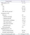
We gathered a total of 321 questionnaires from 25 cardiology centers in Korea. All participants were able to perform or interpret echocardiographic examinations. All participating institutions performed strain echocardiography. Their baseline characteristics are listed in Table 1. In this present study, we included a total of 153 cardiologists, 12 general internists, and 156 echocardiographers. The total participants' career in echocardiography was 7.1 ± 5.8 years in length, and the participants who performed strain echocardiography had longer echocardiographic careers (8.8 ± 5.6 years vs. 4.8 ± 5.3 years, p < 0.001).
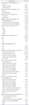
Their awareness of and clinical use of the strain echocardiography are summarized in Table 2. Most of our study participants (97%) were aware of the concept of the strain echocardiography. Most of imaging and heart failure specialists (96%) had high-level knowledge about it.
Of the participants, 186 (58%) performed strain echocardiography. Interestingly, echocardiographers measured strain values more frequently than physicians (85% vs. 32%, p < 0.001). Among cardiologists (n = 164), imaging specialists and heart failure specialists performed strain echocardiography more frequently than other cardiologists or general internists (85% vs. 12%, p < 0.001). The participants used strain echocardiography for clinical and research purposes. However, research purposes seemed to be more frequent. Two-dimensional strain echocardiography was the most commonly used modality in strain echocardiography, and GE was the most frequently used algorithm.
LV was the most commonly used cardiac chamber (99%) for clinical purposes. When RV, left atrium and right atrium were measured, it was usually for research purposes.
Most of the participants did not think LV strain can replace LVEF in their clinical practice, and only 11% of them thought LV strain can be used as LVEF. The most common reason for not performing routine use of strain echocardiography was diversity of strain measurement (65%). Lack of normal reference value and complexity of strain measurement were other reasons. Imaging specialists and heart failure specialists thought diversity is the most common reason for not doing strain echocardiography than other physician (84% vs. 16%, p = 0.008). There was no difference in other reasons.
The most common purpose for clinical and research application of strain echocardiography was the diagnosis of ischemic heart disease (50%). Other purposes of use included prediction of the prognosis of heart failure with reduced ejection fraction (HFrEF), early diagnosis of cardiomyopathy, and prediction of viable myocardium. Imaging specialists and heart failure specialists use strain echocardiography in the diagnosis of ischemic heart disease most commonly. Moreover, they use strain echocardiography in the prediction of viable myocardium, early diagnosis of cardiomyopathy and prediction of prognosis in patients with heart failure frequently. However, there was no statistical difference.
The most common reason of not using strain measurement was no knowledge about the strain echocardiography in general physician (61% vs. 20%, p = 0.002). However, imaging and heart failure specialists did not perform strain measurement because of its complexity and lack of their time (60% vs. 19%, p = 0.002).
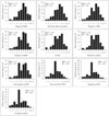
Many participants had a favorable view of the future of strain echocardiography (Fig. 1). They thought strain echocardiography will be most useful in the diagnosis of ischemic heart diseases. Other fields in which strain echocardiography was expected to be useful in included the early diagnosis of cardiomyopathy, the prediction of prognosis in HFrEF and heart failure with preserved ejection fraction, and the prediction of viable myocardium. However, they did not think strain echocardiography will be more useful in the decision of operation time in valvular heart diseases, and application in systemic diseases and prediction of prognosis in valvular heart diseases. Compared to other physicians, imaging specialists and heart failure specialists believed that strain echocardiography will be more useful in almost all fields besides cardiac resynchronization therapy.
Discussion
Most of the participants of this survey had knowledge about strain echocardiography, and all participating institutions performed strain echocardiography in their clinical practice and research fields. Most of them measured LV in their clinical purpose. However, they did not believe that LV strains could replace LVEF for many reasons.
Strain echocardiography is a relatively new echocardiographic modality for measuring myocardial deformations. In Korea, initial introduction of strain echocardiography took place in the research field in the late 1990's. The first Korean article using strain echocardiography was published by Cho et al.16) in 2003. At the time, tissue Doppler imaging was the first modality used to measure myocardial strains. Two-dimensional and three-dimensional strain echocardiography became readily available in current clinical practice with technical improvement. In this survey, all participating institutions performed the strain echocardiography for clinical and research purposes. Interestingly, echocardiographers who measured strains were about 85% of the total number of echocardiographers (132/156). However, only about 33% of the physicians performed strain measurements (54/165). We think that this result was due to the current practice in which echocardiographic examinations are done mostly by echocardiographers, while cardiologists usually confirm the results. However, imaging specialists and heart failure specialists performed strain echocardiography in about 85% in their clinical practice (39/46), so these results should be interpreted with caution.
Interestingly, most of our participants had knowledge of the concept of myocardial strain and strain echocardiography. Moreover, almost all imaging specialists and heart failure specialists thought they have a high level of knowledge. This may be a result of several lectures on strain echocardiography to imaging and heart failure specialists and increased the number of articles using strain echocardiography. However, the percentage of participants who responded that they have no knowledge or moderate level of knowledge was higher in general internists and even in cardiologists other than in imaging and heart failure specialists (53% vs. 4%, p < 0.001). This may be a result of several lectures on strain echocardiography to imaging and heart failure specialists and increased number of articles using strain echocardiography. Education programs showing the strengths and the weaknesses of strain echocardiography will be needed to increase their interest and knowledge, especially for non-imaging and heart failure specialists and general internists. The difference in personal areas of interest can be another reason for this result.
Most of our participants measured LV strain usually for clinical use. This is probably because there are many articles showing LV strain as a good prognostic marker in many cardiovascular diseases.1)2)17) Although there are many study results showing that LV strain has many advantages over LVEF, and many cardiologists are aware of strain echocardiography, the cardiologists generally did not believe that LV strains will replace LVEF in their clinical practice. Because LVEF is the echocardiographic parameter most commonly used to represent LV systolic function, it has been used as a good prognostic parameter in routine clinical practice.1)2) Despite this firm belief, LVEF does have several limitations, and strain echocardiography has been shown to solve these problems in clinical and research fields.6)7) However, only 11% of our participants thought LV strain can be used like LVEF. To change this thinking, more research showing the prognostic significance of LV strains will be necessary. Other studies are also needed to show the strength of strain echocardiography in the detection of subclinical myocardial dysfunction and viable myocardium.
Most participants measured strains of other chambers like RV for research purposes. The main reason for not using RV strains in clinical use was that the estimation of RV systolic function usually depends on visual assessment and strain measurement lacked standardization and normal reference values.18)
Although many still do not think that strain values can be used as an auxiliary indicator of ventricular systolic function, many respondents believed that in the future, strain echocardiography will be more useful in their clinical practice. Imaging specialists had a more favorable outlook on the future of strain echocardiography. To overcome this difference between the high awareness of advantages of strain echocardiography and low application of strain values in actual clinical practice, there are several problems to overcome. The respondents mentioned diversity of strain measurements as the most common reason for not using LV strains instead of LVEF. The second reason was the lack of reference values. To increase the use of the strain echocardiography in clinical practice, it is necessary to use the integrated strain algorithms to solve vendor diversity, as well as to define normal reference values of the strain.
Limitation
This study had several limitations. First of all, this study was based on a questionnaire. Because a survey study allows for generalizable statements about the strain, this study can provide a general sense on the current use of strain echocardiography in the clinical field. However, it may merely represent the data at the present time and give little information on the precise meaning of the data. Moreover, there was room for researcher bias, especially in the preparing of the questionnaire. Secondly, the use of strain echocardiography was dependent on the characteristics of each participating institution. The fact that the institutions that participated in this study were mainly tertiary teaching hospitals may also have influenced the results.
Conclusion
Most of the participants of this study were aware of strain echocardiography, and all institutions performed strain echocardiography for clinical and research purposes. However, the participants did not believe LV strain values could replace LVEF. Because the diversity of strain measurements and lack of normal reference values were common reasons for not using strain echocardiography in the clinical practice, researchers should pay attentions to solve these problems.
 XML Download