

 PDF
PDF ePub
ePub Citation
 Print
Print


Introduction
A prominent crista terminalis is a normal anatomic variant within the right atrium. But, it can be confused with true right atrial mass in transthoracic echocardiogram, especially in patients with embolic events. In this case report, transthoracic echocardiography suggested the presence of a right atrial mass in patients with a pulmonary embolism.
Case
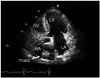
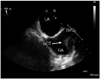
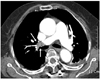
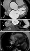
A 73-year-old woman was referred to our hospital for surgery due to tuberculosis spondylitis and concomitant ankylosis. Her blood pressure was 110/70 mmHg, and her electrocariogram showed a normal sinus rhythm with a heart rate of 60 beats per minute. She was underwent surgery without immediate complication. However, twenty days after surgery, the patient complained of dyspnea; the arterial oxygen saturation was decreased to 82%, the D-dimer and B-type natriuretic peptide were increased to 9.73 ug/mL and 2,042 pg/mL, respectively. For evaluation of hypoxia, the transthoracic echocardiogram was perfomed and revealed right side cardiac chamber enlargements without right ventricular dysfunction. The peak pulmonary arterial pressure was increased to 59 mmHg. Furthermore, a 2.2 × 2.4 cm homogenous non-mobile echogenic mass was found within the right atrium (Fig. 1). Because of this finding, we performed a transesophageal echocardiogram that showed the right atrial mass was actually a prominent crista terminalis (Fig. 2). Subsequent chest computed tomography (CT) and magnetic resonance imaging (MRI) revealed findings consistent with a pulmonary embolism (Fig. 3), deep vein thrombosis, and a prominent crista terminalis without any definite right atrial abnormal mass (Fig. 4).
Discussion
The crista terminalis is a fibromuscular ridge derived from the regression of the septum spurium as the sinus venosus is incorporated into the right atrial wall.1) Because of the variation of the regression process, the prominence of the crista terminalis varies widely in adults. Pharr et al.2) reported that if the prominence of the crista terminalis is superior, it can appear as a right atrial mass on the transthoracic echocardiogram. Fig. 2 shows the prominence of the crista terminalis at the superior part of right atrium close to superior vena cava. However, there is no large study on the frequency and characteristics of a prominent crista terminalis with transthoracic echocardiography. Furthermore, all seven patients previously reported in case studies of a prominent crista terminalis were women between the ages of 49 and 77 similar to the case reported here.2-6) Therefore, the age and gender of patients with a prominent crista terminalis appear to be similar to the age and gender of patients with a myxoma. In our case, the patient presented pulmonary embolism after long term immobilization with finding of coincident right atrial mass, which could be misdiagnosed with thrombus or other intra-cardiac mass. Recently, McKay and Thomas4) reported that identification of physiologic structures in the right atrium using additional three dimensional (3D) transthoracic imaging can avoid unnecessary tests such as transesophageal echocardiography or MRI. Thus, we thought that emerging 3D transthoracic imaging could be another alternative modality to differentiate the prominent crista terminalis from the true right atrial mass.
In conclusion, transesophageal echocardiography, CT/MRI, and 3D transthoracic imaging could be used to differentiate a prominent crista terminalis from a true right atrial mass. Understanding of the right atrial anatomy is important to making an accurate diagnosis and to avoid unnecessary additional tests.
 XML Download