

 PDF
PDF ePub
ePub Citation
 Print
Print


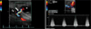
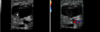
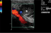
A seventy years-old female complained of a pulsating mass in right inguinal area after elective percutaneous coronary intervention. The size of mass was about 3 cm. Femoral artery US was performed. A 3.2 cm×1.5 cm×15 cm sized pseudoaneurysm which was surrounded by some hematoma was identified. Doppler revealed high velocity blood flow from femoral artery to pseudoaneurysm (Fig. 1). First of all, manual compression was done. Despite of manual compression, there was no definite decrease in size. So, we planned to perform thrombin injection. Thus, total 2000 U of thrombin was injected to pseudoaneurysm, after thrombin injection, with clot formation, blood flow to pseudoaneurysm was nearly disappeared and minimal leak was noted (Fig. 2 and 3).1)2)
Figures and Tables
Fig. 1
Duplex ultrasound image showed 3.2×1.5 cm sized pseudoaneurysm (*) and high velocity flow through a neck (arrow) from femoral artery (FA).
References
1. Middleton WD, Dasyam A, Teefey SA. Diagnosis and treatment of iatrogenic femoral artery pseudoaneurysms. Ultrasound Q. 2005. 21:3–17.
2. Lennox AF, Griffin MB, Chesire NJ, Peters NS, Foale RA, Nicholaides AN. Treatment of an iatrogenic femoral artery pseudoaneurysm with percutaneous duplex-guided injection of thrombin. Circuation. 1999. 100:e39–e41.

 XML Download