

 PDF
PDF ePub
ePub Citation
 Print
Print


Introduction
With recent advances in endovascular treatment, percutaneous transcatheter angioplasty has been increasing for patients with claudication or critical limb ischemia. It can be performed with minimal morbidity and mortality risk. Also, primary stenting in femoropopliteal lesions of intermediate length has recently shown favorable outcomes.1)2)3) However, stent fractures is a concern after bare metal stent implantation . Although incidence of stent fracture vary widely depending on factors such as the treated lesions or stent type, occurrence of stent fractures was reported from 2% to 65%4) which may lead to various complications. While the relationship between stent fractures and restenosis is still controversial, recent studies reported that stent fractures may contribute to late stent failure.5)6)7)
We described a case of stent fracture with complete dislocation combined with recurrent in-stent reocclusion and aneurysm formation in a patient with occlusive disease of the femoropopliteal artery, which was successfully treated with self-expandable Viabahn endoprosthesis.
Case
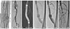
A 71-year old male with multiple comorbidities including coronary and peripheral vascular disease, presented with claudication of left leg of 2 month duration. The patient had a history of diabetes and dyslipidemia. Initial angiography showed total occlusion of distal superficial femoral artery extending to popliteal artery (Fig. 1A). Balloon angioplasty with stent implantation (Absolute Pro, Abbot Vascular, IL, USA) at distal superficial femoral artery was conducted (Fig. 1B). Three months later, he was free of claudication with good patency without aneurysmal changes at previous stent on computed tomography (CT) angiography (Fig. 1C). Seven months later, follow up examination of left femoral stent was performed during percutaneous coronary intervention. Multiple tiny fractures (type II) (Fig. 1D) and 30% neointimal hyperplasia at proximal portion of left superficial femoral stent with aneurysmal change at mid portion of stent (Fig. 1E) were noted; however, the patient was asymptomatic at that time. After six months, he revisited hospital due to recurred left lower extremity claudication. The angiography revealed total occlusion of left distal femoropopliteal artery with previous stent fracture (Fig. 1F). He underwent his second balloon angioplasty for restenosis of the femoral stent and also for popliteal to tibioperoneal trunk.
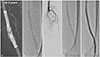
Two years later, on the fourth visit due to recurred claudication (Rutherford class 3), the patient had a cold, pulseless left leg. On angio-CT scan, total destruction in middle part of the left superficial femoral artery stent and aneurysmal change around the lesion (Fig. 2A) were noted. Consistent with CT findings, fluoroscopy showed the stent fracture (type V) (Fig. 2B) with total occlusion of left distal femoral stent (Fig. 2C). After crossing the occlusive lesion with a 0.018 inch guide wire, balloon angiography with 5.0x60 mm balloon was conducted. A 7.0×150 mm self-expandable Viabahn stent (W.L. Gore & Associates, Flagstaff, Ariz, USA) was deployed and adjuvant balloon inflation was done with a 6.0×150 mm balloon (Fig. 2D). The final angiogram showed no residual stenosis and complete exclusion of the pseudoaneurysm (Fig. 2E). There was no complication associated with the procedure, and clinical improvement of symptoms was achieved.
Discussion
Endovascular intervention or bypass surgery is recommended for patients with severe symptoms of peripheral artery disease. Recently, with advances in endovascular treatment, percutaneous transcatheter angioplasty has been increasing for patients with claudication or critical limb ischemia since it can be performed with minimal morbidity and mortality risk. In addition, stent placement has been a standard treatment particularly for medium length femoropopliteal stenosis with evidence from recent randomized trials of higher patency in primary self-expanding nitinol stenting than in provisional stenting.1)2)3)8) However, stent fractures are a concern after bare metal stent implantation. Although the incidences of stent fractures differ according to stent types or lesion characteristics, fractures were reported in 2% to 65% cases, which may lead to serious complications.4) Mechanical factors such as flexion and compression may contribute to stent fracture in peripheral vascular disease. The superficial course of the femoropopliteal arterial segment with crossing of flexion points as well as interaction with the surrounding musculature, potentially expose the vessel to relevant external forces, including compression, torsion, and elongation.9)
Although relation between stent fracture and restenosis is controversial, recent studies reported that stent fractures may contribute to late stent failure5)6)7) and limb-threatening complications.10) In our case, stent placement was performed from distal to proximal popliteal artery. In-stent restenosis was noted although the patient had no symptoms at 7 months after initial interventional treatment. But, at 2 years after initial treatment, he revisited because of recurred severe claudication and previous stent was totally occluded with aneurysmal change around the stent fractures. The cause of aneurysmal change after stent implantation in coronary artery is the excessive use of oversized balloons or high-pressure inflation, resulting in intimal and medial tearing with continuous weakening and stretching of the artery. Another possible mechanism is that a stent fracture with traction between the fragments mechanically breaks down the arterial wall structure and results in aneurysm.11) Although the stent fracture that is related with aneurysmal change in the peripheral arteries have rarely been reported,12)13)14) repeated balloon angioplasty with stent fractures may have attributed to the aneurysmal changes in our patient.
Stent grafts, a combination of a supporting stent structure and a covering graft fabric that were initially used in the treatment of aneurysmal disease have achieved long-term success in the treatment of abdominal aortic and iliac artery aneurysm. Furthermore, stent graft has also been used in the treatment of peripheral arterial occlusive disease such as femoropopliteal arteries, even though maintenance of their patency over the long time remains to be determined. In previous reports, pseudoaneurysm caused by stent fracture was successfully treated by bypass surgery.12)13) Similar to the present case, endovascular stent graft was also performed.14)
In conclusion, we reported a case of femoropopliteal occlusive disease with stent fracture with complete dislocation combined with recurrent in-stent reocclusion and aneurysm formation, which was successfully treated with self-expandable Viabahn endoprosthesis.
 XML Download