

 PDF
PDF ePub
ePub Citation
 Print
Print


Introduction
Coronary aneurysms are identified in 0.15-4.9% of patients undergoing coronary angiography.1) Recently, with the development of non-invasive imaging modalities, including echocardiography, multi-detector computer tomography (MDCT) and magnetic resonance image, the detection of incidental coronary aneurysm is not rare. However, a huge coronary aneurysm, associated with coronary artery fistula, is still uncommon.2-4) Most patients with coronary artery aneurysm are asymptomatic, but occasionally lead to life-threatening conditions, including thrombosis, distal embolization, infection and rupture. Nonetheless, there are still no uniform guidelines for therapeuticmanagement.1)5) We here report a patient who has a huge saccular coronary artery aneurysm with organized thrombi and coronary artery fistulae that was treated by a surgical correction.
Case
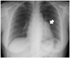
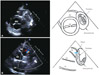
A 48-year-old woman, who had no history of specific conditions, such as Kawasaki's disease or chest trauma, connective tissue disorder was referred to our hospital for the evaluation of an abnormal shadow on the left cardiac border on a chest X-ray film (Fig. 1). She had no subjective symptoms. Her blood pressure was 120/78 mm Hg and pulse rate was 78 beats/min. On physical examination, continuous murmur of a grade II/VI was audible at 4th left intercostal space. The results of other physical examination and blood test were normal. An electrocardiogram showed sinus rhythm and no ST-T wave changes. Transthoracic echocardiography demonstrated a 4.1×4.0 cm round cystic mass with internal echogenecity in the wall, located in contact with upper anterolateral wall of the left ventricle and coronary artery fistula opening to main pulmonary trunk (Fig. 2). Chest MDCT showed a 5 cm saccular aneurysm of proximal left anterior descending artery (LAD) with organized thrombi and multiple fistulae from the conus artery, LAD and aneurysmal sac to the main pulmonary trunk (Fig. 3). Coronary angiography revealed a huge saccular aneurysm at proximal LAD, which was filled with contrast medium in a swirling fashion with slow opacification of distal LAD (thrombolysis in myocardial infarction grade II). Moreover, there were several fistulae from the conus artery of right coronary artery (RCA), proximal LAD, and aneurysmal sac to the pulmonary artery (Fig. 4).
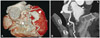
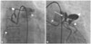
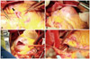
After the diagnosis, she underwent surgery. Median sternotomy was performed. Two vessels from proximal RCA were identified (Fig. 5A). One of them, a fistula from the conus branch to pulmonary artery, were clamped and ligated. After the initiation of cardiopulmonary bypass, the aorta was cross-clamped. When coronary aneurysmal sac was opened, the sac was filled with organized thrombi, which were removed (Fig. 5B and C). Then, the inlet and outlet of aneurysmal sac were ligated and the left internal mammary artery was grafted to the distal LAD. After total bypass, the main pulmonary artery was opened and closed the fistula's opening site, which was observed (Fig. 5D). The postoperative course of the patient was stable and uneventful.
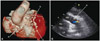
Postoperative MDCT (Fig. 6A) and echocardiography (Fig. 6B), which was performed after 5 days and 7 days, respectively, showed patent bypass graft and successful ligation of aneurysm. However, an abnormal flow at the main pulmonary trunk regarded as a remnant fistula was still visible, although the amount of the abnormal flow was reduced. The patient was discharged, and we have planned regular check-up because the patient was free of symptoms, and the size of fistula was small.
Discussion
Coronary artery aneurysm is defined as a localized dilatation, exceeding the diameter of adjacent normal segment by 1.5 times. Giant coronary artery aneurysms are those with more than 4 cm in diameter.6) Markis et al.7) proposed a classification of aneurysms, according to the morphological feature and the number of affected arteries: type 1 was defined as dilations in all 3 epicardial coronary arteries; type 2, as dilation in 1 blood vessel, only with accompanying stenosis in another coronary artery; and type 3, as the dilation limited only to 1 artery. Coronary aneurysm is most commonly located in the RCA, and then in decreasing order in the left descending artery, the left circumflex artery, and only exceptionally in the left main coronary artery.8)
Coronary-to-pulmonary artery fistula is a congenital heart malformation, and the combination of coronary artery aneurysm and pulmonary artery fistula is very rare. Yu et al.9) reported that coronary artery aneurysm was found in 5.9% of patients with congenital coronary artery fistula, which was found in only 0.2% of patients who had undergone heart surgery.
The majority of the patients with coronary artery aneurysms are asymptomatic, but they may present with angina pectoris, myocardial infarction, sudden death or complications, such as thrombus formation, embolization, fistula formation, rupture, hemopericardium, tamponade, compression of surrounding structure, or congestive heart failure.6)
So far there is no optimal management strategy for patients with giant coronary artery aneurysm. However, depending on the symptoms, etiology and associated lesions, there are a few optional treatments, including medical treatment with anti-platelet agents or anti-coagulation drugs, stent implantation, and surgical exclusion of the aneurysm, using resection or ligation technique.6)10) In our case, 4.1×4.0 cm giant saccular coronary artery aneurysm was located at the proximal left anterior descending coronary artery, and was suspected to be filled with thrombus on transthoracic echocardiography. That could be at high risk of the occlusion of distal LAD and finally myocardial infarction. Also, there is a risk of rupture and tamponade of the giant aneurysm.11)12) Therefore, we decided surgical therapy.
On echocardiogram, after postoperative 7 days, a remnant small fistula from proximal LAD (above aneurysmal sac) across aneurysmal sac to main pulmonary trunk was detected. The optimal therapeutic management of fistula is not well established yet. It is mostly recommended that symptomatic patients (heart failure or ischemia) or patients with large shunts (Qp/Qs>1.5) should be referred for surgery.13)14) On a follow-up echocardiogram, after 3 months, the remnant fistula didn't grow and the value of the shunt (Qp/Qs) was 1.05. We are planning to follow up, on a regular basis, on the patient by echocardiography, even though there is still a remnant small fistula.
A 48-year-old woman has a huge saccular aneurysm with organized thrombi in proximal LAD and coronary artery fistulae from LAD, in addition to the conus branch of RCA to the pulmonary artery. She received surgical treatment, including thrombectomy of aneurysm, ligation of the inlet and outlet of aneurysmal sac, coronary artery bypass graft, and ligation of fistulae. A small remnant fistula, from proximal LAD across aneurysmal sac to pulmonary artery, was observed and will be followed up by echocardiography.
 XML Download