Journal List > Korean Circ J > v.42(11) > 1016960

Lee, Park, Yeo, Sohn, Han, Kim, Kim, Kim, and Chun: Congenital Giant Right Coronary Artery Aneurysm With Fistula to the Coronary Sinus and Persistent Left Superior Vena Cava in an Old Woman

Abstract

The combination of coronary arteriovenous fistula to the coronary sinus (CS), dilatation of the entire length of coronary artery, coronary aneurysm and persistent left superior vena cava (PLSVC) is very rare. We present the case of a 63-year-old female admitted for dyspnea on exertion, orthopnea, and facial edema. Echocardiography detected a giant coronary artery with shunt flow, dilated CS and PLSVC and a coronary angiography reaffirmed these findings. The calculated ratio of pulmonary blood flow to systemic blood flow by cardiac catheterization was 1.53. After multidisciplinary review considering old age, hypoactivity due to underlying Parkinsonism and relatively small amount of shunt flow, medical therapy was chosen. The patient remained asymptomatic for 10 months after discharge without intervention.

Introduction

Congenital coronary arteriovenous fistula (CAVF) has been described to consist of a diagnostic triad: an abnormal localization for a to-and-fro murmur thought to be due to a patent ductus arteriosus; a left to right shunt at the atrial or ventricular level; and a large, tortuous coronary artery on coronary angiography.1) Known complications of CAVF are heart failure, endocarditis, arrhythmias, stroke, myocardial ischemia or myocardial infarction. Many centers have reported surgical corrections or closure of congenital CAVF to prevent the complications.2-7) But choosing an appropriate method of CAVF treatment still remains a controversial issue.8) We report a case of congenital giant right coronary artery which had a fistulous connection to the coronary sinus (CS) and left persistent superior vena cava treated with medical therapy.

Case

A 63-year-old female presented with dyspnea on exertion, orthopnea, and facial edema. She had underlying type 2 diabetes, Parkinsonism, as well as osteoporosis.
At the time of admission, the patient's blood pressure was 100/60 mm Hg, pulse rate was 67/min, and peripheral oxygen saturation was 97%. Physical examination showed mild facial edema without other peripheral limb edemas. The neck vein was slightly engorged. On auscultation, no prominent cardiac murmur was heard.
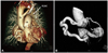
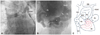
All laboratory values were unremarkable. Chest roentgenography showed an increased cardiothoracic ratio of 0.72 with mild pulmonary congestion. Electrocardiogram showed normal sinus rhythm with left axis deviation. Initial echocardiogram revealed marked dilatation of the right coronary artery (15 mm in diameter) from the initial segment, running along the posterior wall of the left ventricle and terminating around the CS with shunt flow (Fig. 1). The left ventricle was dilated with normal contractility and wall thickness. Computed tomography (CT) angiogram (Fig. 2), following invasive coronary angiogram (Fig. 3B), was performed due to complex anatomy. CT revealed a right coronary artery to CS fistula with persistent left superior vena cava (PLSVC), low lying left innominate vein, and right atrial enlargement possibly due to a left to right shunt.
Cardiac catheterization was performed to determine the amount of shunt flow (Fig. 3A). Cardiac catheterization revealed significant oxygen step-up in the right atrium (RA) {oxygen saturations were as follows: right jugular vein, 51.4%; left subclavian vein, 70.3%; superior vena cava 64.2%; PLSVC (upper) 82.8%; PLSVC (lower) 86.7%; CS ostium 75.7%, high RA 66.4%; mid RA 74.7%; low RA 73.3%; right ventricle 75.8%; pulmonary artery 75.8%, inferior vena cava 61.5%}. The estimated ratio of pulmonary blood flow to systemic blood flow (Qp/Qs) was approximately 1.53.
Based on a multidisciplinary team approach taking into account the patient age and activity level as well as the amount of shunt flow, we concluded to treat her with combination therapy using angiotensin converting enzyme inhibitor and diuretics. After approximately 10 months from the date of discharge, the patient has been doing well without symptom.

Discussion

Coronary arteriovenous fistula is rare cardiac anomaly with a known incidence of 0.3-0.8%.9) Low-pressure structures such as pulmonary artery, superior vena cava, and CS are frequent termination sites of fistulous connections.9) Possible complications include heart failure, angina, endocarditis, arrhythmias, stroke, myocardial ischemia or myocardial infarction due to coronary steal.10) Thus the antiplatelet therapy, especially for patients with distal coronary artery fistulas, abnormally dilated coronary arteries, and prophylactic precautions of subacute bacterial endocarditis, is recommended.8)
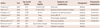
We were able to find eight cases of congenial giant right coronary artery to CS reported in the literature (Table 1).3-6)11-14) Five out of 8 cases were repaired with surgery. Some authors insist, in order to prevent potential complications, that early surgical intervention is the best treatment.5) But surgical intervention for CAVF is still a matter of debate. Some postoperative complications such as myocardial infarction, ischemia, post ligation thrombosis and death have been reported.10)15)16) Liberthson et al.10) reviewed 13 cases of congenital CAVF with surgical therapy and found that postoperative complications increase in patients over 20 years of age. In our case, the patient was treated with loop diuretics and spironolactone for several days after admission after which her facial edema and dyspnea gradually improved. Initially surgical correction or catheter-based closure of fistula was considered, however for symptom improvement, we came across an opportunity to treat with medical therapy. This case suggests that heart failure symptom with Qp/Qs over 1.5 may not be a clear-cut indication for surgery.14)
In conclusion, we report a rare case of CAVF from giant right coronary artery presented with dyspnea and facial edema. To determine the appropriate therapeutic strategy for a rare case such as this one presenting with congenital anomalies, we need to take into account the overall conditions and well-being of the patient.

Figures and Tables

Fig. 1
Transthoracic echocardiography. A: posteriorly angulated apical four-chamber view showed tortuous aneurysmal dilatation of right coronary artery (asterisk) terminating into the coronary sinus (arrow). B: color flow imaging showed shunt flow from the right coronary artery (asterisk) into the coronary sinus (arrow). C: continuous-wave Doppler demonstrated shunt flow throughout whole cardiac cycle with high velocity (4 m/sec). Ao: aorta, CS: coronary sinus, LV: left ventricle, RV: right ventricle.
kcj-42-792-g001
Fig. 2
Volume-rendenered 3-dimensional images of multidetector computed tomography angiogram. A: giant right coronary artery (asterisk); PLSVC (arrow) and left innominate vein ran below the aortic arch (arrow head). B: giant right coronary artery from the initial segement (asterisk); A serpentine coronary artery formed a round mass at the distal end with an opening to the coronary sinus (arrow). An: aneurysm, Ao: aorta, PA: pulmonary artery, PLSVC: persistent left superior vena cava.
kcj-42-792-g002
Fig. 3
Angiography. A: venography with catheterization via coronary sinus to PLSVC (asterisk). B: coronary angiography with pigtail catheter showed the large and tortuous RCA, CS and PLSVC filled with contrast material. Arrow indicates the confluence of PLSVC and RCA to CS. C: drawing elucidating angiographic findings. INN: innominate vein, PLSVC: persistent left superior vena cava, An: aneurysm, Ao: aorta, CS: coronary sinus, IVC: inferior vena cava, PA: pulmonary artery, RA: right atrium, RCA: right coronary artery, RV: right ventricle, SVC: superior vena cava.
kcj-42-792-g003
Table 1
Reported cases of fistulous connection of the right coronary artery to the coronary sinus in adults
kcj-42-792-i001

MR: mitral regurgitation, TR: tricuspid regurgitation

Acknowledgments

This work was supported by 2011 research grant from Pusan National University Yangsan Hospital.

Notes

The authors have no financial conflicts of interest.

References

1. Haller JA Jr, Little JA. Diagnosis and surgical correction of congenital coronary artery-coronary sinus fistula. Circulation. 1963. 27:939–942.
2. Tirilomis T, Aleksic I, Busch T, Zenker D, Ruschewski W, Dalichau H. Congenital coronary artery fistulas in adults: surgical treatment and outcome. Int J Cardiol. 2005. 98:57–59.
3. Ozler A, Tarhan IA, Kehlibar T, et al. Resection of a right coronary artery aneurysm with fistula to the coronary sinus. Ann Thorac Surg. 2008. 85:649–651.
4. Komoda S, Komoda T, Ivanitskaia-Kuehn E, Dreysse S, Pasic M, Hetzer R. Giant aneurysm of the right coronary artery and fistula to the coronary sinus. Gen Thorac Cardiovasc Surg. 2010. 58:78–81.
5. El Watidy AM, Ismail HH, Calafiore AM. Surgical management of right coronary artery-coronary sinus fistula causing severe mitral and tricuspid regurgitation. Interact Cardiovasc Thorac Surg. 2010. 10:110–112.
6. Yu L, Shi E, Gu T. Aneurysmal right coronary with fistula to the coronary sinus combined with severe stenosis of the left anterior descending artery: a snake on the heart. J Thorac Cardiovasc Surg. 2011. 142:937–939.
7. Wilkinson JL. Haemodynamic calculations in the catheter laboratory. Heart. 2001. 85:113–120.
8. Gowda RM, Vasavada BC, Khan IA. Coronary artery fistulas: clinical and therapeutic considerations. Int J Cardiol. 2006. 107:7–10.
9. Yamanaka O, Hobbs RE. Coronary artery anomalies in 126,595 patients undergoing coronary arteriography. Cathet Cardiovasc Diagn. 1990. 21:28–40.
10. Liberthson RR, Sagar K, Berkoben JP, Weintraub RM, Levine FH. Congenital coronary arteriovenous fistula: report of 13 patients, review of the literature and delineation of management. Circulation. 1979. 59:849–854.
11. Yilmaz R, Demirbag R, Gur M. Echocardiographic diagnosis of a right coronary artery-coronary sinus fistula. Int J Cardiovasc Imaging. 2005. 21:649–654.
12. Bittencourt MS, Seltman M, Achenbach S, Rost C, Ropers D. Right coronary artery fistula to the coronary sinus and right atrium associated with giant right coronary enlargement detected by transthoracic echocardiography. Eur J Echocardiogr. 2011. 12:E22.
13. Tekbas G, Onder H, Tekbas E, Yavuz C, Bilici A. Giant right coronary artery and coronary sinus aneurysm due to fistula. Tex Heart Inst J. 2011. 38:314–315.
14. Mitropoulos F, Samanidis G, Kalogris P, Michalis A. Tortuous right coronary artery to coronary sinus fistula. Interact Cardiovasc Thorac Surg. 2011. 13:672–674.
15. Hamada M, Kubo H, Matsuoka H, Kokubu T, Oosuga Y, Joh T. Myocardial infarction complicating surgical repair of left coronary-right ventricular fistula in an adult. Am J Cardiol. 1986. 57:372–374.
16. Loo B, Cox ID, Morgan-Hughes GJ, Marchbank AJ. Thrombotic occlusion of giant circumflex artery aneurysm after ligation of arteriovenous fistula. Circulation. 2010. 122:e447–e448.
TOOLS
Similar articles