

 PDF
PDF ePub
ePub Citation
 Print
Print


Introduction
Angiogenesis is the formation of new blood vessels from preexisting blood vessels, whereas arteriogenesis is the enlarging of existing blood vessels to form collaterals.1) The exact molecular mechanisms that drive neovascularization remain still unclear. Vascular endothelial growth factor-A (or simply VEGF) is a growth factor involved in angiogenesis and arteriogenesis.2)3) VEGF binds to Vascular endothelial growth factor receptor (VEGFR-1/Flt-1) and VEGFR-2 (KDR/Flk-1), but with an approximate 10-fold higher affinity for VE-GFR-1.2)3) Whereas VEGFR-2 mediates almost all of the known cellular responses to VEGF, VEGFR-1 is thought to modulate VEGFR-2 signaling.4) Angiopoietin (Ang)-1 and Ang-2 function as regulators of angiogenesis and arteriogenesis.5-8) Both Angs bind to the extracellular domain of the Tie-2 receptor of endothelial cells.5)6) Whereas Ang-2 destabilizes the vessel to make it sensitive to angiogenic growth factors such as VEGF, Ang-1 promotes vascular stabilization and counteracts VEGF-induced angiogenesis.5)6) Soluble forms of both VEGFR-1 and Tie-2 are produced and secreted into the circulation, where they can bind their respective growth factors and inhibit angiogenesis.9-11) Whereas soluble VEGFR-1 (sFlt-1) is produced by alternative splicing of the VEGFR-1 gene, soluble Tie-2 is the result of proteolytic cleavage from the full-length membrane-bound receptor.9)10) The presence of collateral circulation in patients with acute myocardial infarction (AMI) undergoing primary percutaneous coronary intervention (PCI) has been reported to be associated with smaller infarct size and more preserved left ventricular function.12) Sudden ischemia elicited by myocardial infarction induces a compensatory response to improve myocardial perfusion by angiogenesis and arteriogenesis.13) However, well developed collateral arteries are observed only in a minority of patients at the time of primary PCI.14) Reasons for considerable interindividual variability in the extent of collateral formation remain uncertain. The purpose of the present study was to investigate the relationship between degree of collateral circulation and plasma levels of factors that regulate angiogenesis and arteriogenesis.
Subjects and Methods
Study subjects
A total of 59 consecutive patients (mean age, 59±10 years) with ST-segment elevation myocardial infarct (STEMI) from the registry of the Infarction Prognosis Study, a prospective AMI cohort study at the Severance Cardiovascular Hospital, who underwent successful primary PCI within 12 hours of pain onset during the period between July, 2007 and October 2008 were enrolled in this study. The Infarction Prognosis Study was approved by the hospital institutional review board and performed according to good clinical practice standards. Written informed consent was obtained from each patient before enrollment. The diagnosis of STEMI was based on acute typical chest pain with electrocardiogram findings (ST-segment elevation of ≥0.2 mV in precordial leads and ≥0.1 mV in limb leads), and elevated cardiac troponin T >0.01 ng/mL. All subjects showed a total or subtotal occlusion of the infarct-related artery with Thrombolysis in Myocardial Infarction (TIMI) flow of 0 or 1 on coronary angiography prior to primary PCI. We excluded patients with the following: a past history of old myocardial infarction, having undergone a previous coronary artery bypass surgery, chronic total occlusion, renal failure (serum creatinine >2.0 mg/dL) and presence of significant stenosis in non-infarct related arteries requiring additional revascularization.
Grading of coronary collaterals and primary percutaneous coronary intervention
All patients received loading doses of 250 mg aspirin, 600 mg clopidogrel, and 70 IU/kg of intravenous heparin before primary PCI. Right and left coronary angiography was performed via femoral artery using standard Judkins technique. Degrees of coronary artery stenosis and collateral circulation were estimated visually by 2 independent cardiologists who were blinded to the clinical information of patients. Coronary collateral flow was graded according to the Rentrop scoring system15) as follows: 0, no filling of any collateral vessels; 1, filling of side branches of the artery to be perfused by collateral vessels without visualization of the epicardial segment; 2, partial filling of the epicardial artery by collateral vessels; and 3, complete filling of the epicardial artery by collateral vessels. If collateral circulation to the infarct territory was developed from more than 1 location, the site with the highest grade was taken into account.
All PCIs were performed using a standard technique. A successful PCI was defined as a final TIMI grade 3 flow of the infarct-related artery with <30% residual stenosis. Abciximab was administered during the procedure at the operator's discretion. After PCI, aspirin (100 mg/day) was continued indefinitely, and clopidogrel (75 mg/day) was administered for at least 6 months.
Blood sample collection and laboratory analysis
Blood samples were obtained from an antecubital vein without stasis in all patients immediately after admission in the emergency department and before the administration of heparin. Further blood samples were taken at 24 hours and 48 hours after primary PCI in the same manner from the antecubital vein in all patients. The blood samples, anticoagulated with ethylenediaminetetraacetic acid (EDTA), were immediately centrifuged at 3000 rpm for 10 minutes at 4℃, and an aliquot of the EDTA-plasma was stored at -80℃ until analyzed. Plasma levels of VEGF-A165, sFlt-1, Ang-2, sTie-2 were measured by commercially available enzyme-linked immunosorbent assay kits (R&D Systems, Minneapolis, MN, USA) according to the manufacturer's instruction.
Statistical analysis
Statistical analysis was performed using Statistical Package for the Social Sciences (SPSS) 18.0 for Windows (SPSS Inc., Chicago, IL, USA). Categorical variables were expressed as percentages and compared using chi-square tests between the 2 patient groups. When the expected cell number was <5, Fisher's exact tests were used. Continuous data were expressed as mean±SD. Continuous variables were analyzed using unpaired t-tests and the Mann-Whitney U-test, as appropriate. Correlations between creatine kinase-MB (CK-MB) and growth factor levels were tested using Spearman's rank correlation. The Paired t-test was performed to compare the plasma levels of each angiogenic factor between different time points. For all tests, two-tailed p<0.05 were considered significant.
Results
Clinical and angiographic characteristics
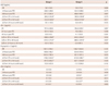
Coronary angiography prior to PCI showed collateral circulation of Rentrop grade 0 in 14, grade 1 in 20, grade 2 in 15, and grade 3 in 10 patients. Patients were divided according to the Rentrop collateral circulation grade into group I (grade 0 or 1, n=34) or group II (grade 2 or 3, n=25). Clinical and angiographic characteristics are summarized in Table 1. Risk factors such as age, gender, hypertension, hypercholesterolemia, and smoking did not differ between the two patient groups. However, diabetes mellitus (41.2% vs.16.0%, p=0.038) were more frequently observed in group I, whereas previous history of angina (18.2% vs. 50%, p=0.011) and multi-vessel disease (47.1% vs. 72.0%, p=0.047) were more frequent in group II.
Previous use of statin, door-to-balloon time, location of infarct-related artery, and serum levels of biomarkers such as cardiac troponin-T, CK-MB, and high sensitivity C-reactive protein did not differ between the two groups. However, group II showed significant lower left ventricular ejection fraction (47.2±9.8% vs. 41.9±9.6%, p=0.049) and a trend toward longer pain-to-balloon time (4.3±2.9 vs. 6.1±6.9, p=0.173) compared to group I.
Vascular endothelial growth factor, soluble VEGF receptor, angiopoietin-2, and sTie2
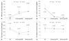
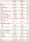
There were increasing plasma levels of VEGF from admission to the emergency room at 24 and 48 hours after PCI. However, there was no statistically significant difference in VEGF plasma levels between the 2 patient groups at any time point (Table 2) (Fig. 1A). Plasma levels of sFlt-1, a VEGF antagonist, were initially elevated, however decreased gradually after PCI (Table 2) (Fig. 1B). Similarly to VEGF, sFlt-1 plasma levels did not differ between the 2 patient groups. In contrast to VEGF, Ang-2 plasma levels were highest on admission and thereafter decreased at 24 and 48 hours after PCI (Table 2) (Fig. 1C). Ang-2 plasma level was significantly higher in group I at 48 hours after PCI than group II (1043.0±785.4 vs. 621.5±381.2 pg/mL, p=0.035). Ang-2 plasma levels on admission (1581.3±594.1 vs. 1364.7±624.5 pg/mL, p=0.181) and 24 hours after PCI (805.8±482.7 vs. 634.6±404.5 pg/mL, p=0.227) tend to be higher in group I than in group II. The difference in plasma levels of VEGF, sFlt-1, and Ang-2 were statistically significant between baseline and 24 hours and between baseline and 48 hours in both groups (Table 2). However, the plasma levels did not change significantly from 24 hours to 48 hours. sTie-2 did not show significant interval changes in plasma concentrations from admission to 24 hours or 48 hours after PCI in group I, but sTie-2 level decreased significantly from baseline to 24 hours or 48 hours in group II (Table 2) (Fig. 1D). However, there was also no statistically significant difference in sTie-2 plasma levels at any time point between the 2 patient groups.
There were no significant correlations between initial CK-MB and initial VEGF levels (r=0.109, p=0.409) and between initial CK-MB and initial Ang-2 levels (r=0.005, p=0.969). Peak CK-MB level did not correlate with any of VEGF (r=0.04, p=0.795) or Ang-2 (r=0.149, p=0.341) levels at 48 hours.
Discussion
Major findings of the present study were the following: 1) Ang-2 plasma levels in patients with STEMI were elevated on admission and fell gradually after primary PCI. 2) Ang-2 plasma levels tend to be slightly higher at baseline and significantly higher at 48 hours after PCI in patients with poor collateral circulation (grade 0/1) than in patients with collateral grade 2/3. 3) VEGF plasma levels were relatively low initially, however, and increased gradually after PCI. 4) sFlt-1, an antagonist of VEGF, showed changes in plasma levels, which were the reverse to those of VEGF. sFlt-1 plasma levels were highest on admission and decreased with time after PCI. 5) VEGF and sFlt-1 levels did not differ within 48 hours according to the extent of collateral circulation. 6) sTie-2 was decreased after PCI only in the patient group with well developed collaterals. However, sTie-2 did not differ significantly between the patient groups during the time course.
Coronary collaterals offer an important alternative source of blood supply when the original vessels occlude due to AMI. In human hearts, an extensive pre-existing collateral network is known to be present.16) The pre-existent collateral vessels usually enlarge upon closure of an epicardial coronary artery, resulting in large collateral conduit arteries. Generally, collateral arteries are evident only in a minority (10-40%) of AMI patients undergoing PCI.14) According to an animal study, de novo collateral artery formation after acute coronary occlusion is usually angiographically notable at least 24 hours after the onset of chest paint.17) Previous studies demonstrated that presence of coronary collateral circulation to the infarct zone was associated with preserved myocardial perfusion and left ventricular (LV) function, reduced in-hospital death, and increased survival of AMI patients.12)18) In our study, patients with collaterals of Rentrop grade 2 or 3 had more frequently previous angina and multi-vessel disease. Diabetes mellitus was associated with poor formation of collateral circulation. These findings were consistent with previous reports. Pre-infarction angina, severity of the infarct-related artery stenosis, right coronary artery occlusion, multi-vessel disease and cholesterol lowering therapy had been suggested as predictors of collateral circulation in patients presenting with AMI.14) However, against our expectation, the patient group with collateral grade 0/1 had higher LV ejection fractions and showed a trend toward lower Killip class compared to the patient group with collateral grade 2/3. This observation may be explained by the higher frequency of multivessel disease and the tendency toward a longer pain-to-balloon time in the patient group with collateral grade 2/3 compared to the patient group with poor collaterals. Multivessel disease usually involves a larger myocardial territory exposed to ischemia. Delayed reperfusion may have resulted in more decreased LV systolic function, however potentially provided more time to recruit collateral vessels.
Angiogenesis, as well as arteriogenesis, is a highly complex process that requires coordinated interaction and cooperation between endothelial cells, extracellular matrix and peri-endothelial cells under the control of growth factors and their receptors. VEGF and Ang-2 are important growth factors involved both in angiogenesis and arteriogenesis.2)3)5-8) sFlt-1 and sTie-2 are circulating decoyed receptors of VEGF and Angio-2, respectively that counteract the actions of the corresponding growth factors. There have been only a little data on the plasma levels of these factors in AMI patients. Ogawa et al.19) reported that serum VEGF level was higher in AMI patients than in patients with stable angina and control healthy subjects. Similarly, Iribarren et al.20) reported that VEGF-A and Ang-2 levels were significantly higher in AMI cases than in controls. Hojo et al.21) performed serial measurements of serum VEGF levels and found that the VEGF levels elevated gradually after the onset of AMI. The peak VEGF level was reached on day 14. Lee et al.22) also investigated serial changes of plasma Ang-1, Ang-2, sTie-2, and VEGF levels from baseline to 18 weeks in AMI. In their study, Ang-1 levels were unchanged from baseline to 6 weeks, but were elevated at 18 weeks. Ang-2 changes followed a biphasic pattern, being higher at baseline and 6 weeks, but lower at 48 hours and 18 weeks. sTie-2 levels increased from baseline and remained elevated in the chronic phase. VEGF peaked at 6 weeks and then decreased toward baseline at 18 weeks. The observation of delayed increase in VEGF levels was supported by the report by Lee et al.23) that VEGF messenger ribonucleic acid (mRNA) expression was only found in human myocardial specimens of "late" infarct (onset of 24 to 120 hours before surgery), whereas Hypoxia-inducible factor (HIF)-1 mRNA was detected in specimens of "acute" infarct (<24 hours before surgery). They suggested that HIF-1 may contribute to the limitation of infarct size by increasing levels of VEGF and thereby promoting angiogenesis. Ang-2 levels are reported to be upregulated by hypoxia.5) Furthermore, previous studies have revealed an ambivalent role of Ang-2 in angiogenesis, depending on the availability of VEGF. For instance, Ang-2 and VEGF act synergistically to produce a stable and functional microvasculature.5)24)25) In the absence of VEGF, however, Ang-2 competitively antagonizes Ang-1-induced Tie-2 phosphorylation and thereby induces vessel regression. The clinical implication of the elevated plasma levels of Ang-2 without rise of VEGF levels at baseline in our study is uncertain. The Ang-2 plasma level tended to be lower prior to primary PCI and was significantly lower at 48 hours after PCI in patients with collaterals of grade 2/3 compared to those with poor collaterals. We assume that the presence of well developed collaterals might not have required higher levels of Ang-2 in response to acute ischemia. However, LV ejection was actually higher in the patient group with collateral grade 0/1 in our study. There was no significant correlation between Ang-2 and CK-MB levels. Furthermore, we cannot explain within the limits of this study why the VEGF levels did not differ between the patient groups. The clinical implications of our findings remain inconclusive. However, to our knowledge, our study is the first clinical study demonstrating the temporal relationships among VEGF, Ang-2, sFlt-1, and sTie-2 in STEMI patients treated with primary PCI. Therefore, our study may deliver the bases for further clinical studies.
Several study limitations need to be acknowledged. First, the sample size was small, which limits statistical power. Therefore, more detailed analysis on various clinical factors or drugs that may have influence the plasma levels of the growth factors could not be performed. Second, the serial measurement of various growth factors was limited to the first 48 hours. Therefore, we cannot rule out the possibility that the plasma levels of growth factors differ between the patient groups later than 48 hours after AMI onset.
In conclusion, our study demonstrated that the plasma levels of Ang-2 and VEGF have different temporal profiles after the onset of AMI. There was a subtle difference in Ang-2 plasma levels according to the extent of collateral circulation development. The presence of collateral circulation in STEMI patients undergoing primary PCI was associated with a lesser rise in plasma levels of Ang-2. VEGF showed a delayed response to acute ischemia compared to Ang-2. The detailed interactions and role of various angiogenic factors in the setting of AMI needs to be investigated in further studies.
 XML Download