

 PDF
PDF ePub
ePub Citation
 Print
Print


Introduction
Hypertension is associated with alterations in the endothelial function of arterial resistance and is often accompanied by severe complications, e.g., stroke, ischemic heart disease and nephrosclerosis, which are associated with vascular damage. The dysfunctional endothelium and resulting structural changes may be responsible for the adverse outcomes of hypertension.1)
Blood pressure (BP) tends to be higher in the early morning hours than at other times of the day, and the nighttime BP is generally 10-20% lower than the daytime BP. A non-dipper rhythm refers to a circadian change in BP in which a nocturnal decrease in the BP is attenuated or absent. Patients with a non-dipper circadian rhythm of the BP have a greater risk of cerebrovascular and cardiovascular complications than those with a dipper circadian rhythm.2)
Endothelial progenitor cells (EPCs) derived from the bone marrow circulate in the peripheral bloodstream and have been implicated in neoangiogenesis after tissue ischemia has occurred, and are candidates for vascular regeneration.3) Thus, circulating EPCs play a key role in the maintenance of endothelial homeostasis and promote vascular repair. They may also have a predictive value for cardiovascular events. A reduced EPC count and associated functional activity have been associated with several cardiovascular risk factors, but their relationship to non-dipper hypertension remains unclear.4-6)
Therefore, we conducted the present study to investigate whether the circulating EPC level was altered in patients with non-dipper hypertension who were at high risk for cardiovascular disease and to evaluate the relationship between the circadian rhythm of the BP and circulating EPC level in patients with essential hypertension.
Subjects and Methods
Subjects
Between December 2007 and May 2008, 47 patients with essential hypertension who were recently identified by outpatient BP measurements and had not previously received antihypertensive therapy were included in this study. After 5 minutes of rest in the sitting position, BP was measured by a calibrated mercury sphygmomanometer, and defined as the average of at least 2 measurements recorded 3 minutes apart. Two patients were excluded because of their refusal, and blood samples were obtained from the remaining 45 patients. Patients with recent cardiovascular events, concomitant malignant diseases or active inflammation were excluded from this study. All individuals were on an unrestricted diet and gave their informed consent according to the protocol approved by the Ethical Committee of our Hospital.
Ambulatory blood pressure monitoring
All patients underwent 24-hour ambulatory BP monitoring (TONOPORT V, GE Marquette, Milwaukee, WI, USA). The patients were divided into two groups, dippers and non-dippers, according to their nocturnal decrease in BP. Patients whose nocturnal decrease in systolic BP was ≥% of the daytime systolic BP were classified as dippers, and those whose nocturnal decrease in systolic BP was <10% of the daytime systolic BP were classified as non-dippers.7)
Isolation of circulating endothelial progenitor cells
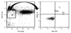
In all participants, the total EPC count was assessed by using an in vitro assay, as described previously.8) In brief, mononuclear cells (MNCs) were obtained from peripheral blood samples (100 µL), and the EPCs were identified by flow cytometry (BD Biosciences, San Jose, CA, USA). Their phenotype was determined by immunohistochemistry after staining with 20 µL fluorescein PE-Cy5-conjugated anti-CD45 monoclonal antibody (Dynona, Korea), 20 µL of fluorescein isothiocyanate (FITC)-conjugated anti-CD34 monoclonal antibody (Dynona, Korea) and 10 µL of PE-conjugated anti-vascular endothelial growth factor receptor 2 (VEGFR2) monoclonal antibody (R&D, Minneapolis, MN, USA), and further incubated in a dark room for 1 hour. After appropriate gating with low cytoplasmic granularity and a low expression of CD45, the number of CD34+VEGFR2+ cells was quantified and expressed as the absolute number of cells per 1×10⁶ peripheral MNCs. The total number of CD45lowCD34+ VEGFR2+ cells was then counted (Fig. 1).
Statistical analysis
All statistical analysis was performed using the Statistical Package for the Social Sciences (SPSS Inc., Chicago, IL, USA) for Windows 12.0. All data are expressed as means±standard deviations. The Student's t-test was used for comparisons of the measured values between the dipper and non-dipper hypertensive patients. A Pearson's correlation coefficient between the number of EPSs and circadian changes in the BP were calculated. A p<0.05 was considered statistically significant.
Results
Subject characteristics
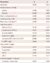
Among the 45 patients with essential hypertension, 20 were classified as dippers (12 men and 8 women; mean age 48±14 years) and 25 as non-dippers (14 men and 11 women; mean age 52±18 years). The baseline characteristics of the dipper and non-dipper hypertensive patients are shown in Table 1. The clinical characteristics and laboratory parameters were similar between the dipper and non-dippers except for the number of lymphocytes (2.5±0.6 vs. 2.1±0.7×10³/uL; p=0.02) and left ventricular ejection fraction (70.8±6.0% vs. 64.8±11.1%; p=0.04).
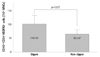
Comparison of differences in the endothelial progenitor cell count between the dippers and non-dippers
Relationship between the endothelial progenitor cell count and parameters
The correlation between the EPC count and parameters is shown in Table 2. The EPC count correlated positively with the number of lymphocytes and the glucose level (r=0.625, p<0.001, and r=0.318, p=0.035, respectively).
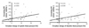
Relationship between the endothelial progenitor cell count and circadian changes in the blood pressure
The correlation between the EPC count and the circadian changes in the BP is presented in Fig. 3. The number of circulating EPCs correlated positively with the circadian changes in the systolic blood pressure and diastolic blood pressure (r=0.435, p=0.003, and r= 0.310, p=0.038, respectively) (Fig. 3).
Discussion
The present study showed that the concentration of the circulating EPCs was significantly reduced in the non-dipper hypertensive patients as compared to the dipper hypertensive patients. Moreover, the circulating EPC count correlated positively with the circadian changes in the systolic and diastolic BPs. To the best of our knowledge, this is the first report to show such a relationship between the EPCs and circadian changes in the BP.
Experimental and clinical studies have suggested that there is an evolving role of the EPCs in neoangiogenesis and rejuvenation of the endothelial monolayer.9)10) Several studies have confirmed that EPCs are derived from the bone marrow, circulate in the peripheral bloodstream and may contribute to new vessel formation by homing in to sites such as ischemic tissues and tumor microenvironments in vitro.11)12) Thus, EPCs would appear to play a key role in the maintenance of endothelial homoeostasis. Recent studies have shown that the EPC count and function can predict the occurrence of cardiovascular events and, most importantly, their imbalance may lead to the development of atherosclerosis.13)
In healthy men, the EPC level may be a surrogate biologic marker for vascular function and cumulative cardiovascular risk. These findings suggest that endothelial injury in the absence of sufficient circulating EPCs may affect the progression of cardiovascular disease.6)
Regarding the relationship between EPCs and cardiovascular events, Schmidt-Lucke et al.14) demonstrated that patients with cardiovascular events have fewer circulating EPCs and that a low number of EPCs independently predicts a higher occurrence of cardiovascular events. Similar results were obtained by Werner et al.5) who found coronary artery disease (CAD) a significant inverse relationship in patients with CAD between the EPC levels and rate of cardiovascular death, revascularization and hospitalization.
Hypertension is one of the most well-known cardiovascular risk factors for target organ damage and cardiovascular events. There is no doubt that the effect of hypertension on the vascular wall is one of the most important determinants of this enhanced risk. With regard to hypertension, it has been shown that patients with CAD have a reduced level and migratory capacity of EPCs, and the latter is mainly influenced by hypertension.
Imanishi et al.15) reported that EPC senescence is accelerated in essential hypertensive patients and positively correlates with the severity of hypertension. Oliveras et al.16) demonstrated that the EPC concentration in the peripheral bloodstream and after in vitro cultures was reduced in patients with refractory hypertension. This decrease was independent of the other risk factors and known determinants of EPCs. This reinforced a likely role of EPCs in atherosclerosis development.
It has been reported that the average lifespan of EPCs is shortened by oxidative stress and regulated by anti-oxidative mechanisms. Angiotensin II receptor antagonists improve EPC dysfunction and increase cardiac c-kit expression through an anti-oxidative mechanism during hypertension. The local renin-angiotensin system induces oxidative stress and regulates EPC function.17-19) In addition, we previously reported that the number of circulating EPCs and flow-mediated vasodilatation (FMD) reduced in vasospastic angina, and statin treatment increased EPC count and FMD.20)
In the present study we showed that the concentration of circulating EPCs is significantly reduced in non-dipper hypertensive patients as compared to dipper hypertensive patients. The reduction in non-dippers may account for the known high cardiovascular risk in these patients. It would be interesting to know the pharmacological and non-pharmacological measures that upregulate EPCs in the early stages of potentially high cardiovascular risk patients to prevent them from developing atherosclerosis and minimize the risk of adverse outcomes.
Our study had several limitations. First, the sample size was small. Second, we could not perform a functional study of the EPCs. Thus, we could not learn the correlation between the EPC functional properties and hypertensive patients. Finally, the specific phenotype of EPCs has not yet been elucidated and there is no defined universal agreement on the characterization of EPCs. Thus, the surface cell markers that we used in our study were not sufficiently precise. However, even considering that all these cells were not true EPCs, they possessed a proven angiogenic potential and may therefore be considered endothelial progenitors in a wide sense.
In conclusion, the present study demonstrated that EPC concentration in the peripheral bloodstream reduced in non-dipper hypertensive patients and circulating EPCs statistically correlated positively with the circadian changes in the BP. However, the clinical significance of the reduced circulating EPCs in non-dippers was unclear. Therefore, these findings need to be confirmed in follow-up studies.

 XML Download