

 PDF
PDF ePub
ePub Citation
 Print
Print


Introduction
Spontaneous chordal rupture is an uncommon clinical complication in patients with hypertrophic cardiomyopathy. To our knowledge, this is the first report of spontaneous chordal rupture in the Korean literature.1-6) Spontaneous chordal rupture results in an altered hemodynamic status secondary to the resolution of left ventricular outflow tract (LVOT) obstruction and the adverse effects of progressive mitral regurgitation.5) Atrial fibrillation is a common complication and contributes to symptomatic deterioration and increased mortality in hypertrophic cardiomyopathy patients.7) This case report described a patient with hypertrophic obstructive cardiomyopathy, spontaneous chordal rupture, and paroxysmal atrial fibrillation. The patient underwent surgical treatment with a mitral valve replacement, septal myectomy, and the Maze procedure.
Case
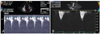
A 69-year-old male patient with chest pain and dyspnea (New York Heart Association class III) was admitted to the hospital. The patient blood pressure was 150/90 mmHg and the heart rate was 65 beats per minute. Electrocardiography revealed atrial fibrillation and negative T waves in the precordial leads. Transthoracic echocardiography revealed asymmetric septal hypertrophy (maximal thickness, 36 mm) and apical hypertrophy (maximal thickness, 18 mm) with a normal ejection fraction (70%). This was compatible with mixed type hypertrophic cardiomyopathy. Severe dynamic LVOT obstruction (LVOT peak velocity=4.6 m/s, peak pressure gradient=85 mmHg) was also noted (Fig. 1A). Moderate eccentric mitral regurgitation was induced by systolic anterior motion of the anterior mitral valve leaflet and prolapse of the posterior leaflet of the medial segment. The effective regurgitant orifice area measured 0.34 cm2, and there was no evidence of chordal rupture. The patient was treated with oral beta blockers and was discharged in stable condition.
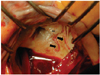
The patient presented to the emergency department 1 year later due to sudden dyspnea (New York Heart Association class IV). Blood pressure was 88/50 mmHg, and electrocardiography demonstrated atrial fibrillation with a rapid ventricular response (145 beats per minute). Chest radiographs revealed cardiomegaly and pulmonary edema of both lung fields. Transesophageal echocardiography revealed flail motion of the medial segments of both mitral leaflets (P3 and A3 segments) with chordal rupture (Fig. 2A). Severe mitral regurgitation occurred secondary to the rupture, and the effective regurgitation orifice area increased to 0.98 cm2 (Fig. 2B). Systolic anterior motion of the mitral leaflet and dynamic LVOT obstruction completely resolved (Fig. 1B). However, aggravated symptoms did not respond to medical management. Concomitant mitral valve replacement, septal myectomy, and the Maze procedure were successfully performed. Spontaneous chordal rupture of the middle segment of the anterior mitral leaflet (A2) and the posteromedial commissure were confirmed during surgery (Fig. 3). Histopathology revealed myxomatous degeneration of the mitral valve without evidence of infective endocarditis. Normal sinus rhythm was maintained postoperatively. Follow-up echocardiography demonstrated a functional prosthetic mitral valve without dynamic LVOT obstruction. The patient was discharged in stable condition with remarkable clinical improvement.
Discussion
Spontaneous chordal rupture is a rare complication of hypertrophic cardiomyopathy. The incidence of simultaneous mitral valve prolapse and hypertrophic cardiomyopathy is approximately 3%. The marked hemodynamic stress resulting from dynamic LVOT obstruction and disrupted mitral valve load-bearing capacity due to myxomatous degeneration contributed to the development of spontaneous chordal rupture8) and acute severe mitral regurgitation in this patient. The presence of chordal rupture was confirmed with transesophageal echocardiography. Chordal rupture was the primary mechanism for progressive mitral regurgitation and acute clinical deterioration in the present report.
Acute chordal rupture of the mitral valve can cause hemodynamic disturbances.1-4)6) A recent report has suggested that chordal rupture in obstructive hypertrophic cardiomyopathy exerts a positive effect on clinical status because the hemodynamic overload associated with mitral regurgitation is counterbalanced by the alleviation of LVOT obstruction.5) Chordal rupture with flailed mitral leaflets was associated with the disappearance of systolic anterior motion of the anterior mitral leaflet and complete relief of severe LVOT obstruction in the present study. This suggested that mitral valve pathology was the key determinant of dynamic LVOT obstruction, irrespective of septal hypertrophy. Additionally, the adverse hemodynamic overload of acute severe mitral regurgitation far outweighed the positive effects of a markedly reduced LVOT pressure gradient in our patient, unlike a previous report.5) This suggested that the clinical manifestations of chordal rupture in obstructive hypertrophic cardiomyopathy are variable in individual patients.
The plausible mechanisms for hemodynamic intolerance in our patient were a decreased threshold for left-sided heart failure due to a decompensated left atrium, pulmonary hypertension, decreased forward cardiac output, and increased numbers of symptomatic episodes of fast atrial fibrillation secondary to atrial pressure overload and subsequent atrial myopathy. In addition, a reduction in diastolic ventricular compliance secondary to myocardial pathology resulted in rapid elevations in left atrial pressure and pulmonary congestion. We performed the Maze operation since atrial fibrillation is poorly tolerated in hypertrophic cardiomyopathy patients and the restoration of sinus rhythm is important for clinical resolution.7)9) Mitral valve replacement was performed instead of valve repair since bi-leaflet prolapse (A2, A3, P3) due to chordal rupture was superimposed on the underlying leaflet redundancy. This was suggestive of myxomatous degeneration. Mitral repair can result in post-operative systolic anterior motion of the anterior mitral leaflet with LVOT obstruction.10) Therefore, prompt surgical approaches including mitral valve replacement, septal myectomy, and the Maze procedure resulted in clinical improvement in the patient.
Transesophageal echocardiography is useful for the detection of recent chordal rupture with severe mitral regurgitation in patients with hypertrophic cardiomyopathy who show sudden hemodynamic decompensation and acute clinical deterioration. Acute mitral regurgitation may cause clinical exacerbation despite improvements in dynamic left ventricular outflow tract obstruction. Therefore, emergency surgery may serve as a therapeutic cornerstone for favorable clinical outcomes.

 XML Download