

 PDF
PDF ePub
ePub Citation
 Print
Print


Various shapes of congenital abnormalities of the menisci have been reported. Among them, the double-layered meniscus is extremely uncommon. Anomalies of the menisci tend to affect the lateral meniscus more commonly and are reportedly more prevalent in Asians than Americans or European populations. There have been a few reports of double-layered lateral meniscus. We report a case of double-layered medial meniscus which have never been reported yet.
CASE REPORT
A 67-year-old female with no history of trauma complained of left knee pain. She had felt pain and discomfort in the left knee for 5 years. On physical examination, she had posteromedial side joint line tenderness, a positive McMurray test on the medial side and normal gait. There were no signs of locking, clicking, ligamentous laxities. She had normal gait, full range of motion, and no giving way sensation.
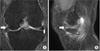
Neurovascular examination was normal. There was no bony anomaly. On coronal, saggital magnetic resonance images posterior portion of radial and horizontal tear of medial meniscus was observed. Double layered medial meniscus was observed anterior to the middle portion of medial meniscus (Fig. 1).
On arthroscopy, complex tear of posterior portion of medial meniscus was observed. The medial meniscus had another meniscus overlying a normal one. The upper meniscus extended from anterior portion to mid portion. Its anterior edge was attached to the lower meniscus. Its periphery was connected to the joint capsule. The posterior portion of upper accessory meniscus was connected to joint capsule (Fig. 2). The accessory meniscus was not mobile on probing. So we think that it is not an origin of symptom, impingement of meniscus.
Only the partial meniscectomy was done on complex tear of posterior portion of medial meniscus. Double-layered medial meniscus was not resected. Postoperatively, ambulation with partial weight bearing was permitted, and the preoperatively pain was allevieadt.
Three months after surgery, the patient could walk freely and returned to normal life without any discomfort.
DISCUSSION
Various anomalies of meniscus have been reported. Among those discoid meniscus was the most common anatomic variant of the meniscus.1) Other less common anomalies include incomplete discoid meniscus, Wrisberg meniscus, ring-shaped meniscus, accessory meniscus, congenital separation meniscus, congenital partial deficiency and absence of the menisci.2)
Double-layered meniscus have been reported with a prevalence of 0.06% to 0.09%.3) Suzuki et al.1) reported two cases of double-layered lateral meniscus with one meniscus overlying the other. Double-layered medial meniscus was an extremely rare anatomical abnormality more than lateral meniscus.
Double-layered lateral meniscus was thin, mobile in the previous case report.14) But in this case, double-layered medial meniscus was cord like round shape. And it was not mobile. We think that it was not an origin of pain. These findings were different from the double-layered lateral meniscus which was reported previously. 1235)
Komatsu et al.6) reported double-layered medial meniscus. The anterior portion of the medial meniscus had two layers, attached to the anterior surface of the tibia. In this case the anterior portion of the medial mesniscus had two layers but it was attached to lower normal medial meniscus.
Lee and Min7) reported abnormal band of the lateral meniscus. It was characteristic that upper abnormal band was loose and serpentine. In this case upper accessory meniscus was not loose, nor mobile.
Regarding the differentiation of double layered meniscus from a horizontal tear, the cleavage of a horizontal tear is sharp margin, irregular in shape.3) In this case, the margin was smooth, regular in both upper and lower menisci. In addition, there were no problems with respect to the volume of the residual meniscus suggesting that the anomaly was not due to a horizontal tear (Fig. 2). Based on these findings, we concluded that this anomaly was different from acquired changes induced by degeneration or trauma.
Menisci differentiate directly from blastemal cells connected to the capsule.48) The causes of such variations are multifactorial, including congenital and developmental influences, but clinical presentations, pathology, and epidemiology of variations, altered biomechanics of medial compartment are still unclear.2)
After surgery, the patient had no discomfort or pain. We think that double-layered medial meniscus was not the cause of pain. It is unclear how double-layered medial meniscus influences knee joint biomechanics and also double-layered medial meniscus can be a cause of meniscus tear.
Double-layered medial meniscus was an extremely rare anatomical abnormality. We thought that abnormal band of meniscus,7) double-layered meniscus,123) separated meniscus6) were similar meniscus abnormalites. But they were a little different. It was unclear that morphological classification, its characteristics. It is expected to clarify its characteristics, altered biomechanics, influence to original meniscus and causes of double-layer medial meniscus.

 XML Download