

 PDF
PDF ePub
ePub Citation
 Print
Print


Abstract
Purpose
We compared the ocular aberration and clinical outcome between different aspheric intraocular lenses (IOL) in both eyes.
Methods
This prospective randomized controlled study was comprised of patients with bilateral cataract who received two different aspheric IOLs implanted in both eyes: negatively aspheric Tecnis® ZCB00 and spherically neutral Akreos® MI60. Total and corneal aberrations computed by Wavescan® and Pentacam® were assessed at 6 months to investigate the effects of the IOL's spherical aberration on the eye and to analyze the incidence and degree of posterior capsule opacification. By using spherical aberration of the cornea and the IOLs, values calculated via Ray-tracing software and Wavescan® were compared. Total spherical aberration was analyzed by the MATLAB program and converting the pupil size to 6.0, 4.5, 3.0 mm.
Results
A total of 25 patients were included. Regarding pre-operative corneal aberration, ZCB00 group was 0.232 ± 0.119 µm while MI60 group was 0.240 ± 0.117 µm, and there was no difference between the two IOLs. At 6 months after total ocular spherical aberration, MI60 group (pupil size 6.0 mm; 0.296 ± 0.097 µm, 4.5 mm; 0.094 ± 0.032 µm, 3.0 mm; 0.019 ± 0.006 µm) had more positive values than ZCB00 group (pupil size 6.0 mm; 0.051 ± 0.105 µm, 4.5 mm; 0.009 ± 0.034 µm, 3.0 mm; 0.002 ± 0.007 µm) (p < 0.001). When calculated using the ray tracing method, based on the results after surgery, MI60 group's total spherical aberrations were higher than ZCB00 group. However, from 1 month to 6 months after surgery, the uncorrected distance visual acuity, spherical equivalent and posterior capsule opacification showed no differences between the two IOLs.
Conclusions
In eyes with aspheric IOLs with negative spherical aberration, spherical aberration was lower than spherically neutral aspheric IOLs. Regarding postoperative visual acuity, spherical equivalent and posterior capsule opacification, there were no significant differences between the two groups.
Figures and Tables
Figure 1
Posterior capsule opacification (PCO) severity grading with POCOman software. (A) Retroilluminated slit lamp photographs 6 months postoperatively. (B) PCO score with POCOman software. The severity score is calculated according to the formula: (Blue × 1 + Yellow × 2 + Red × 3)/total area.
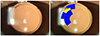
Figure 2
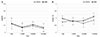
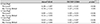
Visual acuity and manifest refraction at 6 months postoperatively. (A) Uncorrected distance visual acuity (UDVA, logMAR). (B) Spherical equivalent. There was no difference in UDVA and spherical equivalent between the 2 intraocular lens groups.

References
1. Werner L, Olson RJ, Mamalis N. New technology IOL optics. Ophthalmol Clin North Am. 2006; 19:469–483.
2. Holladay JT, Piers PA, Koranyi G, et al. A new intraocular lens design to reduce spherical aberration of pseudophakic eyes. J Refract Surg. 2002; 18:683–691.
3. Rawer R, Stork W, Spraul CW, Lingenfelder C. Imaging quality of intraocular lenses. J Cataract Refract Surg. 2005; 31:1618–1631.
4. Guirao A, Redondo M, Geraghty E, et al. Corneal optical aberrations and retinal image quality in patients in whom monofocal intraocular lenses were implanted. Arch Ophthalmol. 2002; 120:1143–1151.
5. Lombardo M, Lombardo G. Wave aberration of human eyes and new descriptors of image optical quality and visual performance. J Cataract Refract Surg. 2010; 36:313–331.
6. Williams D, Yoon GY, Porter J, et al. Visual benefit of correcting higher order aberrations of the eye. J Refract Surg. 2000; 16:S554–S559.
7. Montés-Micó R, Ferrer-Blasco T, Cerviño A. Analysis of the possible benefits of aspheric intraocular lenses: review of the literature. J Cataract Refract Surg. 2009; 35:172–181.
8. Nochez Y, Favard A, Majzoub S, Pisella PJ. Measurement of corneal aberrations for customisation of intraocular lens asphericity: impact on quality of vision after micro-incision cataract surgery. Br J Ophthalmol. 2010; 94:440–444.
9. Nanavaty MA, Spalton DJ, Boyce J, et al. Wavefront aberrations, depth of focus, and contrast sensitivity with aspheric and spherical intraocular lenses: fellow-eye study. J Cataract Refract Surg. 2009; 35:663–671.
10. McKelvie J, McArdle B, McGhee C. The influence of tilt, decentration, and pupil size on the higher-order aberration profile of aspheric intraocular lenses. Ophthalmology. 2011; 118:1724–1731.
11. Sasaki K, Sakamoto Y, Shibata T, et al. Measurement of postoperative intraocular lens tilting and decentration using Scheimpflug images. J Cataract Refract Surg. 1989; 15:454–457.
12. Bender L, Spalton DJ, Uyanonvara B, et al. POCOman: new system for quantifying posterior capsule opacification. J Cataract Refract Surg. 2004; 30:2058–2063.
13. Werner W, Roth EH. Image properties of spherical as aspheric intraocular lenses. Klin Monbl Augenheilkd. 1999; 214:246–250.
14. Mester U, Dillinger P, Anterist N. Impact of a modified optic design on visual function: clinical comparative study. J Cataract Refract Surg. 2003; 29:652–660.
15. Kershner RM. Retinal image contrast and functional visual performance with aspheric, silicone, and acrylic intraocular lenses. Prospective evaluation. J Cataract Refract Surg. 2003; 29:1684–1694.
16. Pandita D, Raj SM, Vasavada VA, et al. Contrast sensitivity and glare disability after implantation of AcrySof IQ Natural aspherical intraocular lens: prospective randomized masked clinical trial. J Cataract Refract Surg. 2007; 33:603–610.
17. Tzelikis PF, Akaishi L, Trindade FC, Boteon JE. Spherical aberration and contrast sensitivity in eyes implanted with aspheric and spherical intraocular lenses: a comparative study. Am J Ophthalmol. 2008; 145:827–833.
18. Rocha KM, Soriano ES, Chalita MR, et al. Wavefront analysis and contrast sensitivity of aspheric and spherical intraocular lenses: a randomized prospective study. Am J Ophthalmol. 2006; 142:750–756.
19. Guirao A, Tejedor J, Artal P. Corneal aberrations before and after small-incision cataract surgery. Invest Ophthalmol Vis Sci. 2004; 45:4312–4319.
20. Oshika T, Sugita G, Miyata K, et al. Influence of tilt and decentration of scleral-sutured intraocular lens on ocular higher-order wavefront aberration. Br J Ophthalmol. 2007; 91:185–188.
21. Taketani F, Matuura T, Yukawa E, Hara Y. Influence of intraocular lens tilt and decentration on wavefront aberrations. J Cataract Refract Surg. 2004; 30:2158–2162.
22. Suh Y, Oh C, Kim HM. Comparison of the long-term clinical results of hydrophilic and hydrophobic acrylic intraocular lenses. Korean J Ophthalmol. 2005; 19:29–33.
23. Heatley CJ, Spalton DJ, Kumar A, et al. Comparison of posterior capsule opacification rates between hydrophilic and hydrophobic single-piece acrylic intraocular lenses. J Cataract Refract Surg. 2005; 31:718–724.
24. Auffarth GU, Golescu A, Becker KA, Völcker HE. Quantification of posterior capsule opacification with round and sharp edge intraocular lenses. Ophthalmology. 2003; 110:772–780.
25. Morgan-Warren PJ, Smith JA. Intraocular lens-edge design and material factors contributing to posterior-capsulotomy rates: comparing Hoya FY60aD, PY60aD, and AcrySof SN60WF. Clin Ophthalmol. 2013; 7:1661–1667.
26. Vock L, Menapace R, Stifter E, et al. Posterior capsule opacification and neodymium:YAG laser capsulotomy rates with a round-edged silicone and a sharp-edged hydrophobic acrylic intraocular lens 10 years after surgery. J Cataract Refract Surg. 2009; 35:459–465.
27. Vasavada AR, Raj SM, Shah A, et al. Comparison of posterior capsule opacification with hydrophobic acrylic and hydrophilic acrylic intraocular lenses. J Cataract Refract Surg. 2011; 37:1050–1059.
28. Nanavaty MA, Spalton DJ, Gala KB, et al. Fellow-eye comparison of posterior capsule opacification between 2 aspheric microincision intraocular lenses. J Cataract Refract Surg. 2013; 39:705–711.
29. Li Y, Wang J, Chen Z, Tang X. Effect of hydrophobic acrylic versus hydrophilic acrylic intraocular lens on posterior capsule opacification: meta-analysis. PLoS One. 2013; 8:e77864.
30. Nagata T, Minakata A, Watanabe I. Adhesiveness of AcrySof to a collagen film. J Cataract Refract Surg. 1998; 24:367–370.
31. Dorey MW, Brownstein S, Hill VE, et al. Proposed pathogenesis for the delayed postoperative opacification of the hydroview hydrogel intraocular lens. Am J Ophthalmol. 2003; 135:591–598.
32. Marcos S, Barbero S, Jiménez-Alfaro I. Optical quality and depth-of-field of eyes implanted with spherical and aspheric intraocular lenses. J Refract Surg. 2005; 21:223–235.
33. Rocha KM, Soriano ES, Chamon W, et al. Spherical aberration and depth of focus in eyes implanted with aspheric and spherical intraocular lenses: a prospective randomized study. Ophthalmology. 2007; 114:2050–2054.
34. Jeong JH, Kim MK, Wee WR, Lee JH. Comparison of optical performances in eyes implanted with aspheric and spherical intraocular lenses after cataract surgery. J Korean Ophthalmol Soc. 2010; 51:1445–1452.
 XML Download