

 PDF
PDF ePub
ePub Citation
 Print
Print


Abstract
Purpose
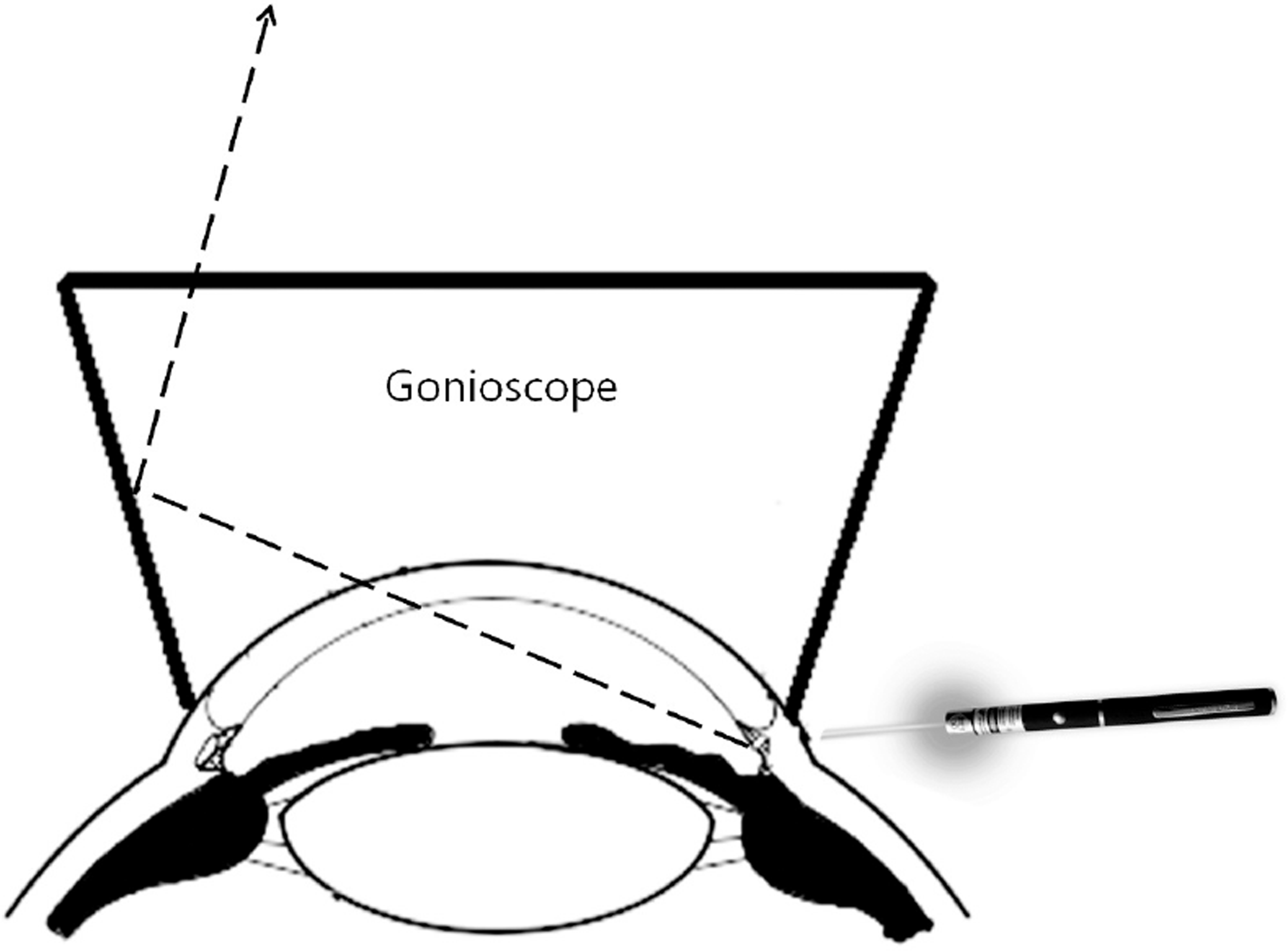
To introduce a novel adjuvant technique to locate cyclodialysis cleft using a laser pointer in a gonioscopic view.
Case summary
A 36-year-old man complaining of blurred vision in his left eye after blunt trauma 2 weeks prior was referred to our hospital. Gonioscopy showed a cyclodialysis cleft from 3 to 4 o’clock and fundus revealed hypotonic maculopathy. After the failure of medical treatment, we tried various interventions such as injection of viscoelastic agent into the anterior chamber and intravitreal gas tamponade with transconjunctival cryotherapy. Since those were not successful, we decided to treat the patient with direct cyclopexy. For the preoperative localization of the cleft, we tried a new technique that uses a laser pointer. On gonio-scopic examination, an assistant shot the laser toward the limbal area where the suspicious cleft was located. We were able to precisely locate the cyclodialysis cleft if the laser pointer light was seen through the cleft in the gonioscopic view. With the aid of a laser a pointer, the cleft was successfully closed.
References
1. Ceruti P, Tosi R, Marchini G. Gas tamponade and cyclocryotherapy of a chronic cyclodialysis cleft. Br J Ophthalmol. 2009; 93:414–6.

2. Ioannidis AS, Barton K. Cyclodialysis cleft: causes and repair. Curr Opin Ophthalmol. 2010; 21:150–4.
3. Ioannidis AS, Bunce C, Barton K. The evaluation and surgical management of cyclodialysis clefts that have failed to respond to conservative management. Br J Ophthalmol. 2014; 98:544–9.
4. Nolan W. Anterior segment imaging: ultrasound biomicroscopy and anterior segment optical coherence tomography. Curr Opin Ophthalmol. 2008; 19:115–21.
5. Monaco WA, Barker FM 2nd. Laser hazards and safety. Optom Clin. 1995; 4:1–15.
Figure 1.
Localization of the cyclodialysis cleft with the laser pointer, during gonioscopic examination. (A) Laser pointer light in normal gonioscopic view. (B) Macular wrinkling was shown at fundus photography. (C) The novel method with laser pointer was used for cyclodialysis cleft localisation. (D) After direct cyclopexy, the cyclodialysis cleft was closed. (E) The laser pointer that was used to locate of small cyclodialysis clefts.

 XML Download