

 PDF
PDF ePub
ePub Citation
 Print
Print


Abstract
Purpose
To evaluate the association between body mass index (BMI) and visual field (VF) progression in normal tension glaucoma (NTG) patients.
Methods
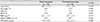
We reviewed the medical records of 78 eyes of 78 NTG patients who were treated with eye drops for more than 18 months. Age, sex, existence of hypertension (HTN), diabetes mellitus (DM), refractive error, baseline intraocular pressure (IOP), IOP reduction ratio, baseline VF indices including mean deviation (MD) and pattern standard deviation, VF progression rate (MD slope, dB/year), number of eye drops, and BMI were analyzed. The progression of VF was determined by glaucoma change probability analyses (STATPAC 2) using a Humphrey field analyzer.
Results
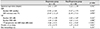
The mean follow-up in consecutive eyes was 4.4 ± 2.7 years. A total of 18 eyes showed progression and 60 eyes did not. The VF progression rate (p < 0.001) and number of eye drops (p = 0.024) showed statistical differences, but age, sex, existence of HTN and DM, refractive error, baseline IOP, IOP reduction ratio, baseline VF index, and BMI did not show a statistical difference between the two groups (all, p > 0.05). However, multiple linear regression analyses showed that a lower BMI was significantly associated with faster VF progression in the progression group (β = 0.078; standard error = 0.030; p = 0.027).
Figures and Tables
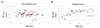
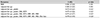
Figure 1
Correlations between body mass index (BMI) and the visual field (VF) progression in the progression and non-progression groups. (A) The progression group. (B) The non-progression group (line: linear regression line). MD = mean deviation.
References
1. Shields MB. Normal-tension glaucoma: is it different from primary open-angle glaucoma? Curr Opin Ophthalmol. 2008; 19:85–88.
2. Kim CS, Seong GJ, Lee NH, et al. Prevalence of primary open-angle glaucoma in central South Korea the Namil study. Ophthalmology. 2011; 118:1024–1030.
3. Caprioli J, Spaeth GL. Comparison of visual field defects in the low-tension glaucomas with those in the high-tension glaucomas. Am J Ophthalmol. 1984; 97:730–737.
4. Tokunaga T, Kashiwagi K, Tsumura T, et al. Association between nocturnal blood pressure reduction and progression of visual field defect in patients with primary open-angle glaucoma or normal-tension glaucoma. Jpn J Ophthalmol. 2004; 48:380–385.
5. Marcus DM, Costarides AP, Gokhale P, et al. Sleep disorders: a risk factor for normal-tension glaucoma? J Glaucoma. 2001; 10:177–183.
6. Ong K, Farinelli A, Billson F, et al. Comparative study of brain magnetic resonance imaging findings in patients with low-tension glaucoma and control subjects. Ophthalmology. 1995; 102:1632–1638.
7. Klaver JH, Greve EL, Goslinga H, et al. Blood and plasma viscosity measurements in patients with glaucoma. Br J Ophthalmol. 1985; 69:765–770.
8. Tripolino C, Irace C, Carallo C, et al. Body fat and blood rheology: Evaluation of the association between different adiposity indices and blood viscosity. Clin Hemorheol Microcirc. 2017; 65:241–248.
9. Polsky S, Ellis SL. Obesity, insulin resistance, and type 1 diabetes mellitus. Curr Opin Endocrinol Diabetes Obes. 2015; 22:277–282.
10. Kwagyan J, Tabe CE, Xu S, et al. The impact of body mass index on pulse pressure in obesity. J Hypertens. 2005; 23:619–624.
11. Drager LF, Togeiro SM, Polotsky VY, Lorenzi-Filho G. Obstructive sleep apnea: a cardiometabolic risk in obesity and the metabolic syndrome. J Am Coll Cardiol. 2013; 62:569–576.
12. Zang EA, Wynder EL. The association between body mass index and the relative frequencies of diseases in a sample of hospitalized patients. Nutr Cancer. 1994; 21:247–261.
13. Pasquale LR, Willett WC, Rosner BA, Kang JH. Anthropometric measures and their relation to incident primary open-angle glaucoma. Ophthalmology. 2010; 117:1521–1529.
14. Ramdas WD, Wolfs RC, Hofman A, et al. Lifestyle and risk of developing open-angle glaucoma: the Rotterdam study. Arch Ophthalmol. 2011; 129:767–772.
15. Kim KE, Kim MJ, Park KH, et al. Prevalence, awareness, and risk factors of primary open-angle glaucoma: Korea National Health and Nutrition Examination Survey 2008-2011. Ophthalmology. 2016; 123:532–541.
16. Gasser P, Stumpfig D, Schötzau A, et al. Body mass index in glaucoma. J Glaucoma. 1999; 8:8–11.
17. Kim M, Jeoung JW, Park KH, et al. Metabolic syndrome as a risk factor in normal-tension glaucoma. Acta ophthalmol. 2014; 92:e637–e643.
18. Kim YH, Jung SW, Nam GE, et al. High intraocular pressure is associated with cardiometabolic risk factors in South Korean men: Korean National Health and Nutrition Examination Survey, 2008-2010. Eye (Lond). 2014; 28:672–679.
19. Kim YJ, Chun YS, Lee MY, et al. Association of IOP with systemic factors in a Korean cohort. Optom Vis Sci. 2015; 92:1182–1188.
20. Kim MJ, Park KH, Kim CY, et al. The distribution of intraocular pressure and associated systemic factors in a Korean population: the Korea National Health and Nutrition Examination Survey. Acta ophthalmol. 2014; 92:e507–e513.
21. Jang HD, Kim DH, Han K, et al. Relationship between intraocular pressure and parameters of obesity in Korean adults: The 2008-2010 Korea National Health and Nutrition Examination Survey. Curr Eye Res. 2015; 40:1008–1017.
22. Lee JY, Kim TW, Kim HT, et al. Relationship between anthropometric parameters and open angle glaucoma: The Korea National Health and Nutrition Examination Survey. PloS One. 2017; 12:e0176894.
23. Chan TCW, Bala C, Siu A, et al. Risk factors for rapid glaucoma disease progression. Am J Ophthalmol. 2017; 180:151–157.
24. Lyu IJ, Lee JM, Kee C. Risk factors for rapid visual field progression in normal-tension glaucoma. J Korean Ophthalmol Soc. 2012; 53:996–1001.
25. Kim NY, Seo HR, Rho SH. The relationships among delayed recovery in finger temperature, nocturnal dip, and glaucoma progression. J Korean Ophthalmol Soc. 2015; 56:70–79.
26. Kim HA, Han K, Lee YA, et al. Differential association of metabolic risk factors with open angle glaucoma according to obesity in a Korean population. Sci Rep. 2016; 6:38283.
 XML Download