

 PDF
PDF ePub
ePub Citation
 Print
Print


Abstract
Purpose
To help predict the severity of ocular complications in orbital blunt trauma by analyzing clinical features of ocular complications and orbital wall fracture.
Methods
A retrospective survey consisting of 169 eyes of 168 patients with orbital blunt trauma who visited Kosin University Gospel Hospital was performed. The patients' age, gender, height, weight, cause of accident, and ocular complications were investigated. The patients were imaged using computer tomography and divided into two groups according to whether they had orbital wall fracture: the orbital contusion group and the orbital wall fracture group. Variables of orbital wall fracture, including the location, length, width, and area were measured using computer tomography in the orbital wall fracture group.
Results
The incidence of ocular complication was 27 of 67 eyes (40.2%) in the orbital wall fracture group and 75 of 102 eyes (73.5%) in the orbital contusion group; ocular complications were found significantly more often in the orbital contusion group than in the orbital wall fracture group (p < 0.001). Among causes of the accident, ‘flying object’ showed the highest incidence of ocular complication at 40 of 48 eyes (85.1%). In the orbital wall fracture group, there was no significant difference in orbital fracture location, length, width or area between the ocular complication group and the non-ocular complication group.
References
1. Kreidl KO, Kim DY, Mansour SE. Prevalence of significant abdominal sequelae in blunt orbital trauma. Am J Emerg Med. 2003; 21:525–8.
2. He D, Blomquist PH, Ellis E 3rd. Association between ocular abdominal and internal orbital fractures. J Oral Maxillofac Surg. 2007; 65:713–20.
3. Lee TS, Kellman R, Darling A. Crumple zone effect of nasal cavity and paranasal sinuses on posterior cranial fossa. Laryngoscope. 2014; 124:2241–6.

4. Mellema PA, Dewan MA, Lee MS, et al. Incidence of ocular injury in visually asymptomatic orbital fractures. Ophthal Plast Reconstr Surg. 2009; 25:306–8.
5. Cook T. Ocular and periocular injuries from orbital fractures. J Am Coll Surg. 2002; 195:831–4.
6. Shin JW, Lim JS, Yoo G, Byeon JH. An analysis of pure blowout fractures and associated ocular symptoms. J Craniofac Surg. 2013; 24:703–7.
7. Lee JS, Lee H, Shin H, et al. Significant intraocular sequelae in abdominalal blunt trauma and their association with blowout fracture. J Korean Ophthalmol Soc. 2012; 53:613–7.
8. Waterhouse N, Lyne J, Urdang M, Garey L. An investigation into the mechanism of orbital blowout fractures. Br J Plast Surg. 1999; 52:607–12.
9. Nagasao T, Miyamoto J, Jiang H, et al. Interaction of hydraulic and buckling mechanisms in blowout fractures. Ann Plast Surg. 2010; 64:471–6.
10. Rhee JS, Kilde J, Yoganadan N, Pintar F. Orbital blowout fractures: experimental evidence for the pure hydraulic theory. Arch Facial Plast Surg. 2002; 4:98–101.
Figure 1.
The measurement of variables of orbital wall fracture. (A-C) Location and anterior to posterior length of orbital wall fracture. For example, Distance A to B: (The number of computed tomography [CT] cross section between A and B) × (CT slice thickness [2 mm or 3 mm]). (A) Both lacrimal crests (red arrows). (B) Starting point of orbital wall fracture (red arrow). (C) End point of orbital wall fracture (red arrow). (D, E) Longest length of orbital wall fracture. (D) Length of medial orbital wall fracture on coronal section. (E) Length of inferior orbital wall fracture on coronal section. (F-I) Orbital wall fracture areas. To calculate the area of the orbital wall fracture, we measured lengths of the orbital wall fracture in all CT section, and then multiplied it by CT slice thickness (2 mm or 3 mm). The sum of these values is orbital fracture area. For example; 4.04 × 3 + 12.49 × 3 + 6.61 × 3 + 7.68 × 3 = Orbital fracture area.
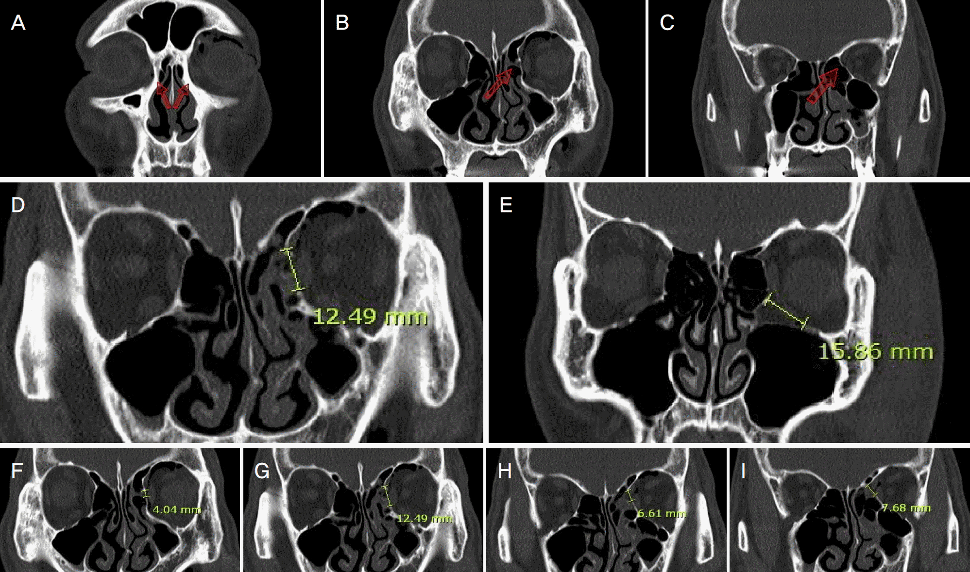
Table 1.
Comparison of base line characteristics of patients between ocular complication group and no ocular complication group
| Baseline characteristics | Total | Complications* | No complication† | p-value |
|---|---|---|---|---|
| Number of eye | 169 | 102 | 67 | |
| Sex (male/female) | 140/29 | 83/19 | 57/10 | |
| Laterality (OD/OS) | 75/94 | 41/61 | 34/33 | |
| Age (years) | 36.1 ± 17.8 | 35.7 ± 17.0 | 36.5 ± 19.1 | 0.770‡ |
| Height (cm) | 168.0 ± 9.6 | 169.3 ± 9.5 | 166.3 ± 9.7 | 0.160‡ |
| Weight (kg) | 67.12 ± 12.8 | 67.1 ± 11.1 | 67.0 ± 14.8 | 0.963‡ |
Table 2.
Comparing frequency of ocular complication between orbital contusion group and orbital wall fracture group
| Ocular complication | Orbital contusion* (n = 102) | Orbital wall fracture† (n = 67) | p-value§ |
|---|---|---|---|
| Commotio retinae | 40 (40.4) | 19 (28.4) | 0.112 |
| Traumatic iritis | 47 (46.1) | 13 (19.4) | <0.001 |
| Hyphema | 20 (19.6) | 2 (2.9) | 0.002 |
| Angle recession | 5 (4.9) | 2 (2.9) | 0.541 |
| Retinal hemorrhage | 5 (4.9) | 2 (2.9) | 0.541 |
| Cyclodialysis | 4 (3.9) | 0 (0) | 0.101 |
| Traumatic cataract | 3 (2.9) | 0 (0) | 0.157 |
| Vitreous hemorrhage | 1 (0.9) | 0 (0) | 0.416 |
| Lens dislocation | 0 (0) | 0 (0) | Not comparable |
| Retinal detachment | 0 (0) | 0 (0) | Not comparable |
| Eyeball rupture | 0 (0) | 0 (0) | Not comparable |
| The number of complicated eyes | ‡75 (73.5) | 27 (40.2) |
Table 3.
Variables of orbital wall fracture
| Variables of orbital wall fracture | Complications group* | No complication† | p-value# |
|---|---|---|---|
| Number | 26 | 40 | |
| AP length from lacrimal crest to starting point of fracture (mm)‡ | 11.3 ± 3.7 | 11.4 ± 2.5 | 0.949 |
| AP length (mm)† | 21.8 ± 5.6 | 20.6 ± 5.6 | 0.381 |
| Longest length in coronal plane CT (mm)§ | 17.5 ± 4.1 | 17.1 ± 4.3 | 0.649 |
| Fracture area (mm2)Π | 323.6 ± 155.1 | 287.6 ± 133.1 | 0.335 |
Table 4.
Comparing cause of accident between ocular complications group and no ocular complication group
| Cause of accident | Complications group* | No complication† | Total |
|---|---|---|---|
| Flying object | 40 (85.1) | 7 (14.9) | 47 |
| Falling down | 22 (45.8) | 26 (54.2) | 48 |
| Assault | 34 (54.8) | 28 (45.2) | 62 |
| Traffic accident | 6 (50) | 6 (50) | 12 |
| Total | 102 (60.3) | 67 (39.7) | 169 |
 XML Download