

 PDF
PDF ePub
ePub Citation
 Print
Print


Abstract
Purpose
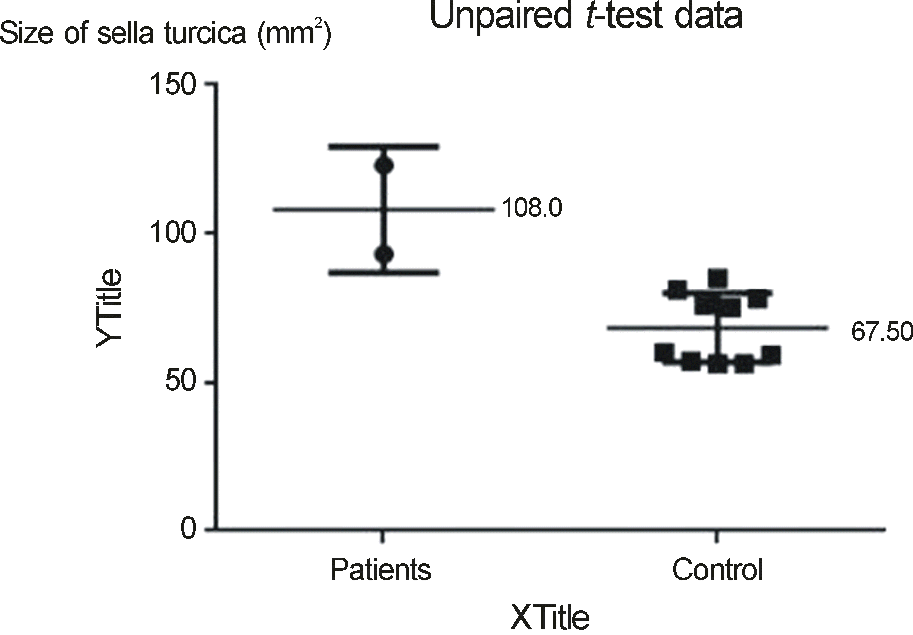
In this study we evaluated the hypothesis that sella turcica enlarged in size due to increased intracranial hypertension by measuring the sella turcica area using magnetic resonance imaging (MRI) in patients with increased intracranial hypertension and compared to normal controls.
Methods
Brain magnetic resonance (MR) midsagittal images of patients diagnosed with pseudotumor cerebri from 2005 to 2012 at Dankook University Hospital and 10 normal controls who had no overt signs or symptoms of neurological disease and had nor-mal gadolinium-enhanced MR examination of brain were compared. The area of the sella turcica was measured by the dou-ble-blind method using Dicomworks v 1.3.5b (Philippe Puech and Loic Boussel, Freeware, France). Statistical analysis was con-ducted using GraphPad Prism (GraphPad Software, Inc., USA) and Mann-Whitney U-test.
References
1. Brodsky MC, Vaphiades M. Magnetic resonance imaging in pseu-dotumor cerebri. Ophthalmology. 1998; 105:1686–93.

2. Park JW, Cha SH, Song GS, Choi CH. Pseudotumor cerebri asso-ciated with polycythemia vera. J Korean Neurosurg Soc. 2003; 34:162–4.
3. Takanashi J, Suzuki H, Nagasawa K, et al. Empty sella in children as a key for diagnosis. Brain and Development. 2001; 23:422–3.
4. Messina D, Bono F, Fera F, et al. Empty sella and bilateral trans-verse sinus stenosis predict raised intracranial pressure in the ab-sence of papilloedema. J Neurol. 2006; 253:674–6.
5. Wessel K, Thron A, Linden D, et al. Pseudotumor cerebri: clinical and neuroradiological findings. Eur Arch Psychiatry Neurol Sci. 1987; 237:54–60.
6. Kaufman B. The “empty” sella turcica–a manifestation of the intrasellar subarachnoid space. Radiology. 1968; 90:931–41.
7. Friedman DI, Jacobson DM. Diagnostic criteria for idiopathic in-tracranial hypertension. Neurology. 2002; 59:1492–5.
8. Yuh WT, Zhu M, Taoka T, et al. MR imaging of pituitary morphol-ogy in idiopathic intracranial hypertension. J Magn Reson Imaging. 2000; 12:808–13.
9. Davis PC, Hoffman JC Jr, Spencer T, et al. MR imaging of pituitary adenoma: CT, clinical, and surgical correlation. AJR Am J Roentgenol. 1987; 148:797–802.
10. Weisberg LA, Numuguchi Y. Neuroimaging in neuroendocrine diseases. Neurol Clin. 1986; 4:783–800.
11. Hwang TN, Rofagha S, McDermott MW, et al. Sunken eyes, sag-ging brain syndrome: bilateral enophthalmos from chronic intra-cranial hypotension. Ophthalmology. 2011; 118:2286–95.
12. Thurtell MJ, Bruce BB, Newman NJ, Biousse V. An update on idi-opathic intracranial hypertension. Rev Neurol Dis. 2010; 7:e56–68.
Figure 1.
(A) T1-weighted mid-sagittal image of the sella turcica and pituitary gland from a normal subject. (B) Drawing showing the dimensions that were measured to determine the opening (solid line) and contour (dotted line) of the sella turcica. In this example, the sella area = 93 mm.
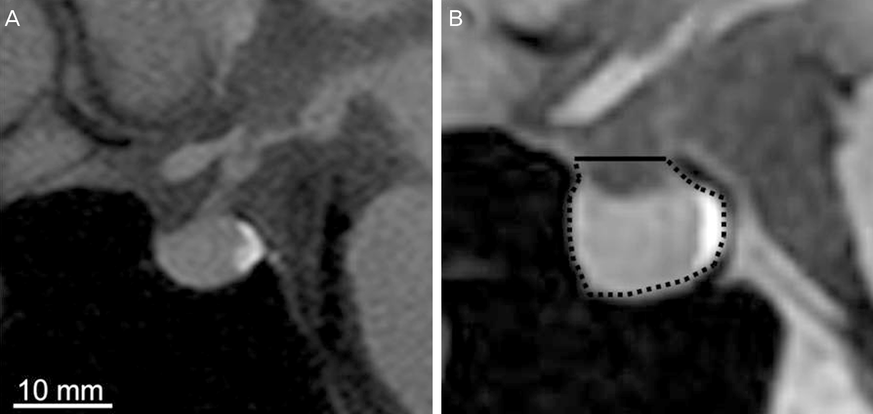
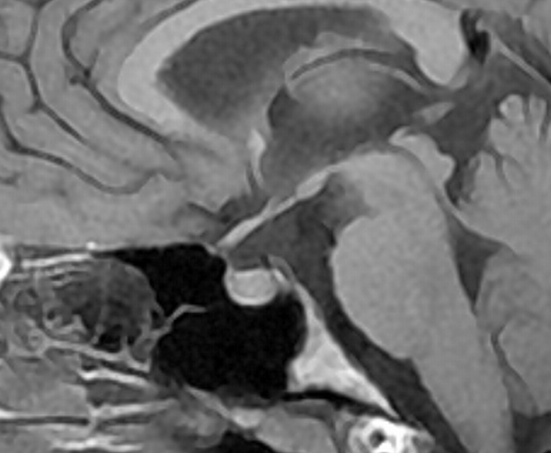
Figure 2.
Tl-weighted mid-sagittal image of 17-year-old wom-an patient showed partially empty sella (turcica area: 93 mm2).
 XML Download