Abstract
Purpose
To compare higher-order aberrations (HOAs) and visual acuity after wavefront-guided and wavefront-optimized laser keratorefractive surgery.
Methods
This retrospective study consisted of consecutive cases of eyes that underwent wavefront-guided (VISX S4 CustomVue®) or wavefront-optimized (WaveLight® EX500) laser assisted in situ keratomileusis (LASIK) or photorefractive keratectomy (PRK). Preoperative and postoperative uncorrected visual acuity (UCVA), best corrected visual acuity (BCVA), manifest refraction spherical equivalent (MRSE), and preoperative and 3 month postoperative HOAs were compared.
References
1. Netto MV, Dupps W Jr, Wilson SE. Wavefront-guided ablation: evidence for efficacy compared to traditional ablation. Am J Ophthalmol. 2006; 141:360–8.

2. Thibos LN, Applegate RA, Schwiegerling JT, Webb R. Standards for reporting the optical aberrations of eyes. J Refract Surg. 2002; 18:S652–60.

3. Netto MV, Ambrósio R Jr, Shen TT, Wilson SE. Wavefront analysis in normal refractive surgery candidates. J Refract Surg. 2005; 21:332–8.

4. Yoon G, Macrae S, Williams DR, Cox IG. Causes of spherical aberration induced by laser refractive surgery. J Cataract Refract Surg. 2005; 31:127–35.

5. Roberts C. Biomechanics of the cornea and wavefront-guided laser refractive surgery. J Refract Surg. 2002; 18:S589–92.

6. Ahn SM, Seok SS, Park CY. Considering spherical aberration in choosing the wavefront map for laser vision correction. J Korean Ophthalmol Soc. 2011; 52:147–56.

7. Miraftab M, Seyedian MA, Hashemi H. Wavefront-guided vs wavefront-optimized LASIK: a randomized clinical trial comparing contralateral eyes. J Refract Surg. 2011; 27:245–50.

8. Buzzonetti L, Iarossi G, Valente P, et al. Comparison of wavefront aberration changes in the anterior corneal surface after laser-as-sisted subepithelial keratectomy and laser in situ keratomileusis: preliminary study. J Cataract Refract Surg. 2004; 30:1929–33.

9. Chalita MR, Chavala S, Xu M, Krueger RR. Wavefront analysis in post-LASIK eyes and its correlation with visual symptoms, refraction, and topography. Ophthalmology. 2004; 111:447–53.

10. Perez-Straziota CE, Randleman JB, Stulting RD. Visual acuity and higher-order aberrations with wavefront-guided and wavefront-optimized laser in situ keratomileusis. J Cataract Refrac Surg. 2010; 36:437–41.

11. Padmanabhan P, Mrochen M, Basuthkar S, et al. Wavefront-guided versus wavefront-optimized laser in situ keratomileusis: contralateral comparative study. J Cataract Refract Surg. 2008; 34:389–97.

12. Racine L, Wang L, Koch DD. Size of corneal topographic effective optical zone: comparison of standard and customized myopic laser in situ keratomileusis. Am J Ophthalmol. 2006; 142:227–32.

13. Zhou C, Chai X, Yuan L, et al. Corneal higher-order aberrations after customized aspheric ablation and conventional ablation for myopic correction. Curr Eye Res. 2007; 32:431–8.

14. Brint SF. Higher order aberrations after LASIK for myopia with al-con and wavelight lasers: a prospective randomized trial. J Refract Surg. 2005; 21:S799–803.

15. Stonecipher KG, Kezirian GM. Wavefront-optimized versus wavefront-guided LASIK for myopic astigmatism with the ALLEGRETTO WAVE: three-month results of a prospective FDA trial. J Refract Surg. 2008; 24:S424–30.
16. Mrochen M, Kaemmerer M, Seiler T. Clinical results of wavefront-guided laser in situ keratomileusis 3 months after surgery. J Cataract Refract Surg. 2001; 27:201–7.

17. Hong JT, Lee JE, Kim JY, et al. Clinical results of wavefront-guided LASIK. J Korean Ophthalmol Soc. 2010; 51:1438–44.

18. Lee SM, Lee MJ, Kim MK, et al. Comparison of changes in high-er-order aberrations between conventional and wavefront-guided PRK. J Korean Ophthalmol Soc. 2007; 48:1028–35.
19. Moshirfar M, Espandar L, Meyer JJ, et al. Prospective randomized trial of wavefront-guided laser in situ keratomileusis with the CustomCornea and CustomVue laser systems. J Cataract Refract Surg. 2007; 33:1727–33.

20. Awwad ST, Bowman RW, Cavanagh HD, McCulley JP. Wavefront-guided LASIK for myopia using the LADAR CustomCornea and the VISX CustomVue. J Refract Surg. 2007; 23:26–38.

21. Tran DB, Shah V. Higher order aberrations comparison in fellow eyes following intraLase LASIK with wavelight allegretto and customcornea LADArvision4000 systems. J Refract Surg. 2006; 22:S961–4.

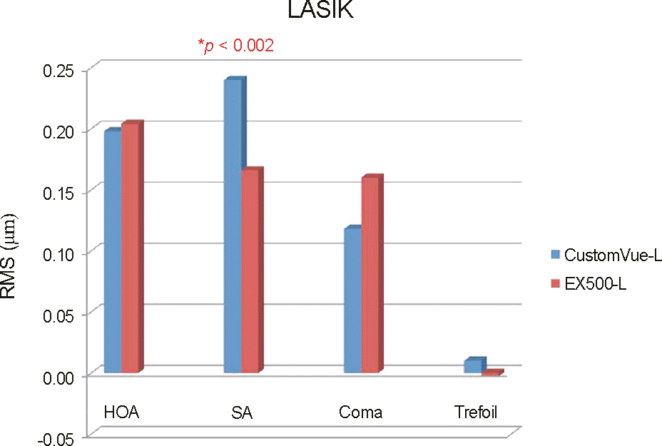
Figure 1.
Postoperative changes in HOAs by group in LASIK. HOA = higher-order aberration; SA = spherical aberration.

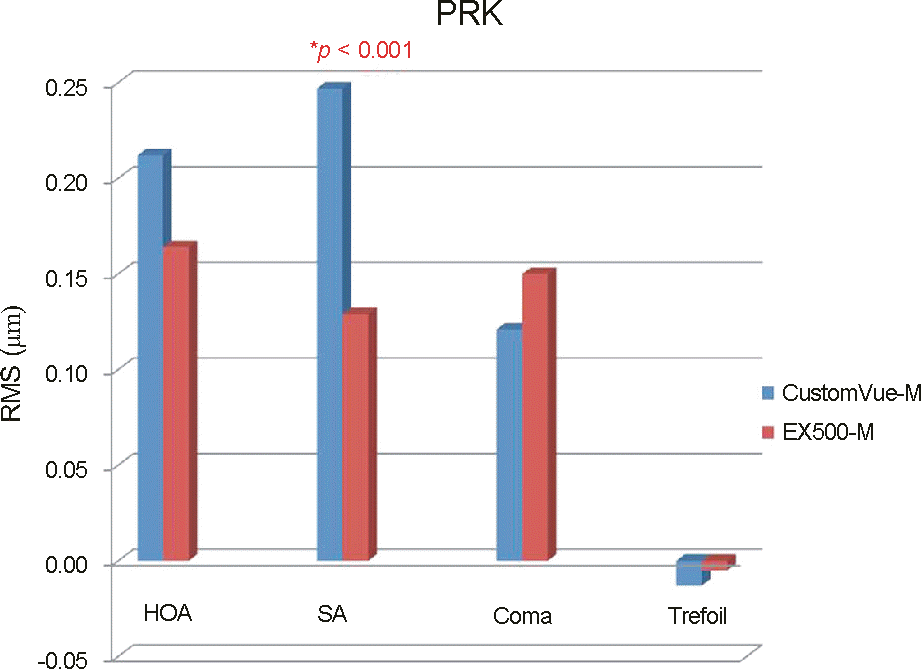
Figure 2.
Postoperative changes in HOAs by group in PRK. HOA = higher-order aberration; SA = spherical aberration.

Table 1.
Patient demographics of LASIK group
|
Group |
p-value | ||
|---|---|---|---|
| CustomVue | EX500 | ||
| Age (years) | 31.59 ± 9.09 | 28.71 ± 6.57 | 0.33 |
| UCVA (log MAR) | 1.15 ± 0.37 | 1.15 ± 0.35 | 0.76 |
| MRSE (D) | - 3.64 ± 1.75 | - 3.60 ± 1.60 | 0.92 |
Table 2.
Patient demographics of PRK group
|
Group |
p-value | ||
|---|---|---|---|
| CustomVue | EX500 | ||
| Age (years) | 29.27 ± 5.87 | 26.00 ± 6.09 | 0.11 |
| UCVA (log MAR) | 1.52 ± 0.34 | 1.40 ± 0.37 | 0.77 |
| MRSE (D) | -4.02 ± 1.69 | -4.39 ± 1.68 | 0.28 |
Table 3.
Preoperative HOAs of LASIK group
| HOA RMS | CustomVue | EX500 | p-value |
|---|---|---|---|
| Total HOA (μm) | 0.39 ± 0.15 | 0.37 ± 0.12 | 0.48 |
| Spherical aberration | 0.11 ± 0.14 | 0.08 ± 0.13 | 0.09 |
| Coma | 0.21 ± 0.13 | 0.20 ± 0.11 | 0.51 |
| Trefoil | 0.20 ± 0.11 | 0.20 ± 0.10 | 0.97 |


 PDF
PDF ePub
ePub Citation
 Print
Print


 XML Download