

 PDF
PDF ePub
ePub Citation
 Print
Print


Abstract
Purpose
To evaluate the clinical manifestations of double elevator palsy and describe the surgery outcomes in patients.
Methods
We performed a retrospective chart review of all patients who were treated surgically for double elevator palsy between 1999 and 2012 at Yonsei University, Severance Hospital in Seoul, Korea.
Results
Overall, 15 subjects (7 males and 8 females) with a mean age of 14.6 years (range, 3-40 years) underwent their first surgery during the study period. All patients received inferior rectus recession as a primary procedure. Nine patients (60.0%) underwent a secondary procedure which included 4 cases of horizontal muscle transposition, 2 cases of correction of exotropia, and 3 cases of correction of hypotropia and exotropia simultaneously. The mean preoperative hypotropia was decreased from 29.9 ± 8.4 prism diopter (PD) to 4.7 ± 5.3 PD postoperatively. Mean follow-up period was 40.9 ± 48.2 months. Seven patients (46.7%) underwent eyelid surgery for true ptosis. At last follow-up, a majority of patients showed mild or no amblyopia.
Conclusions
Primary inferior rectus recession and additive secondary horizontal muscle transposition surgery was effective in treatment of double elevator palsy. The clinical manifestations and surgical outcomes of monocular elevation deficiency in the present study can help in the treatment of Korean patients.
References
1. Knapp P. The surgical treatment of double-elevator paralysis. Trans Am Ophthalmol Soc. 1969; 67:304–23.
2. Rose LV, Elder JE. Management of congenital elevation deficiency due to congenital third nerve palsy and monocular elevation deficiency. Clin Experiment Ophthalmol. 2007; 35:840–6.

3. Yurdakul NS, Ugurlu S, Maden A. Surgical treatment in patients with double elevator palsy. Eur J Ophthalmol. 2009; 19:697–701.
4. Mcneer KW, Jampolsky A. Double elevator palsy caused by anomalous insertion of the inferior rectus. Am J Ophthalmol. 1965; 59:317–9.
5. Ford CS, Schwartze GM, Weaver RG, Troost BT. Monocular elevation paresis caused by an ipsilateral lesion. Neurology. 1984; 34:1264–7.
6. Muñoz M, Page LK. Acquired double elevator palsy in a child with a pineocytoma. Am J Ophthalmol. 1994; 118:810–1.
7. Olson RJ, Scott WE. Dissociative phenomena in congenital monocular elevation deficiency. J AAPOS. 1998; 2:72–8.
8. Bagheri A, Sahebghalam R, Abrishami M. Double elevator palsy, subtypes and outcomes of surgery. J Ophthalmic Vis Res. 2008; 3:108–13.
9. Pediatric Eye Disease Investigator Group. The clinical profile of moderate amblyopia in children younger than 7 years. Arch Ophthalmol. 2002; 120:281–7.
10. Metz HS. Double elevator palsy. Arch Ophthalmol. 1979; 97:901–3.
11. Scott WE, Jackson OB. Double elevator palsy: the significance of inferior rectus restriction. Am Orthopt J. 1977; 27:5–10.
12. Kim JH, Hwang JM. Congenital monocular elevation deficiency. Ophthalmology. 2009; 116:580–4.
13. Ziffer AJ, Rosenbaum AL, Demer JL, Yee RD. Congenital double elevator palsy: vertical saccadic velocity utilizing the scleral search coil technique. J Pediatr Ophthalmol Strabismus. 1992; 29:142–9.
14. Burke JP, Ruben JB, Scott WE. Vertical transposition of the horizontal recti (Knapp procedure) for the treatment of double elevator palsy: effectiveness and long-term stability. Br J Ophthalmol. 1992; 76:734–7.
15. Caldeira JA. Vertical transposition of the horizontal rectus muscles for congenital/early onset “acquired” double elevator palsy: a retrospective long term study of 10 consecutive patients. Binocul Vis Strabismus Q. 2000; 15:29–38.
16. Kocak-Altintas AG, Kocakkkk-Midillioglu I, Dabil H, Duman S. Selective management of double elevator palsy by either inferior rectus recession and/or knapp type transposition surgery. Binocul Vis Strabismus Q. 2000; 15:39–46.
17. Metz HS. Double elevator palsy. J Pediatr Ophthalmol Strabismus. 1981; 18:31–5.
18. Williams C, Northstone K, Harrad RA, et al. Amblyopia treatment outcomes after preschool screening v school entry screening: observational data from a prospective cohort study. Br J Ophthalmol. 2003; 87:988–93.
19. Jewell G, Reeves B, Saffin K, Crofts B. The effectiveness of vision screening by school nurses in secondary school. Arch Dis Child. 1994; 70:14–8.
20. Lennerstrand G, Jakobsson P, Kvarnström G. Screening for ocular dysfunction in children: approaching a common program. Acta Ophthalmol Scand Suppl. 1995; 214:26–38. discussion 39-40.
Figure 1.
Preoperative and postoperative visual acuity. Mild amblyopia better than 6/12, moderate 6/12–6/30, and severe amblyopia worse than 6/30–3/60 (derived from Pediatric Eye Disease Investigator Group definitions). (A) Total (B) patients who underwent their strabismus surgery under the age of 6 (C) patients who underwent their strabismus surgery over the age of 6.
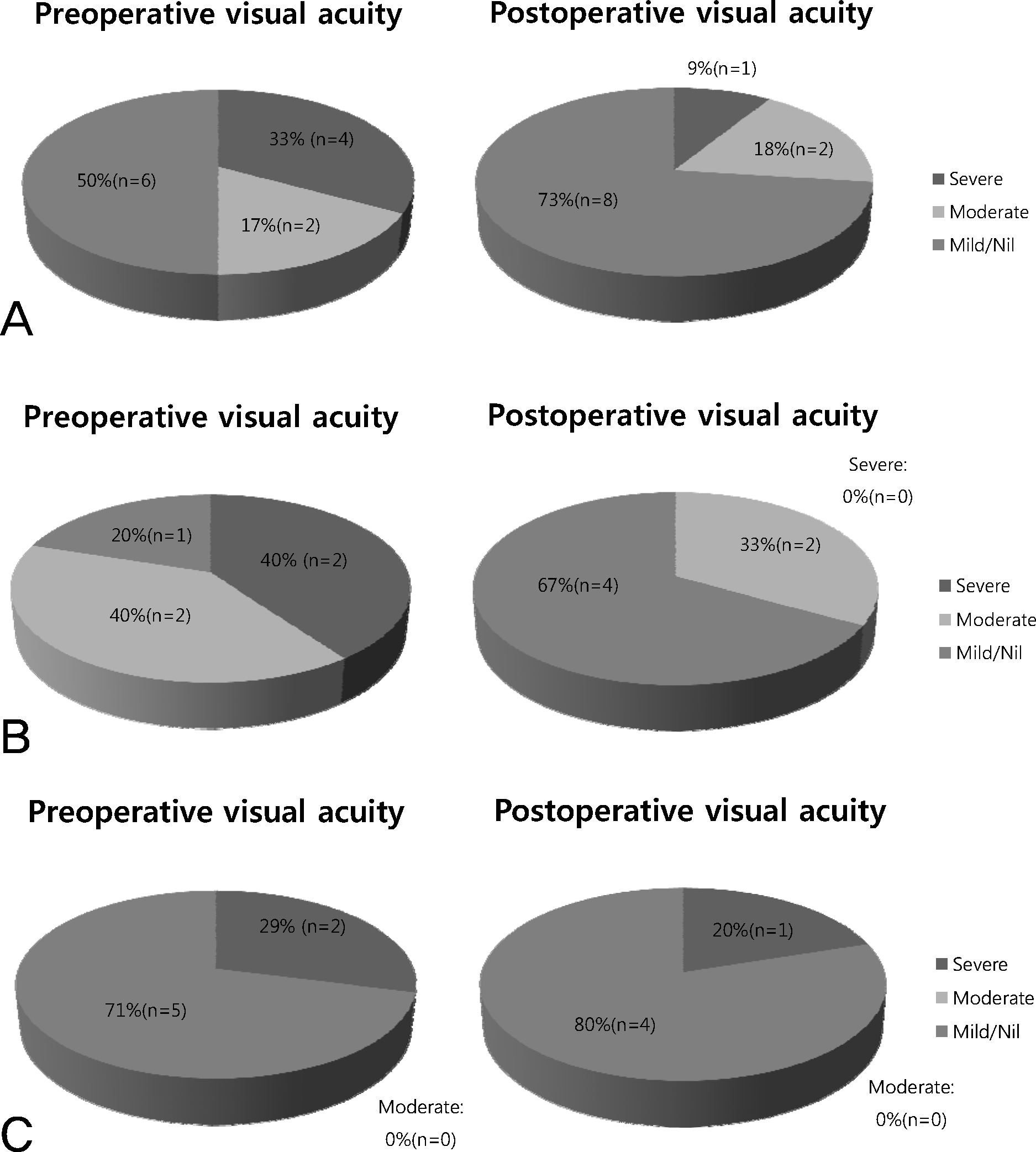
Table 1.
Background and demographic data of patients with double elevator palsy
Table 2.
Strabismus and ptosis procedures performed in group 1 (negative traction test) and group 2 (positive traction test)
| Patient//sex/age | Pre-op deviation (PD) | Forced duction test | Initial surgery (mm) | Second pre-op deviation (PD) | Second surgery (mm) | Final deviation (PD) | Length of follow-up (months) | Ptosis | Interval between strabismus op and ptosis op |
|---|---|---|---|---|---|---|---|---|---|
| Group 1 | |||||||||
| 1/F/40 | 30 LHoT | Negative | IR recess 7.0 | − | − | 16 LHoT | 32 | − | − |
| 2/M/18 | 30 RHoT | Negative | IR recess 5.0 SO tenotomy | − | − | 10 XT | 183 | − | − |
| 3/F/10 | 14 RHoT 14 XT | Negative | IR recess 6.0 LR recess 6.5 | 14 RHoT | Horizontal Hummelsheim op | 3 RHoT | 57 | True ptosis | 6 months after initial op |
| 4/M/4 | 35 LHoT | Negative | IR recess 7.0 | − | − | Ortho | 12 | True ptosis | 10 months after initial op |
| 5/F/29 | 40 LHoT | Negative | IR recess 6.0 | 40 XT | LR recess 10.0 MR resection 7.0 | 3 LHoT | 18 | True ptosis | 1 year after initial op |
| 6/F/1 | 30 RHoT | Negative | IR recess 6.0 | 14 RHoT 10 XT | MR half tendon up transposition LR recess 2.0 | 8 RHoT | 21 | Pseudoptosis | − |
| 7/F/30 | 20 LHoT | Negative | IR recess 6.0 | − | − | 7 LHoT 7 XT | 12 | − | − |
| 8/M/4 | 45 LHoT | Negative | IR recess 6.0 | 30 LHoT | Knapp op | Ortho | 57 | True ptosis | 7 months after second op |
| 9/F/5 | 35 RHoT | Negative | IR recess 6.0 | 16 RHoT 14 XT | LR half tendon up transposition MR resection 5.0 | 6 RHoT | 12 | True ptosis | 10 months after second op |
| 10/F/3 | 25 LHoT 20 XT | Negative | IR recess 5.0 | 12 LHoT 14 XT | SR resection 4.5 LR recess 5.5 | 6 LHoT 5 XT | 21 | − | − |
| Group 2 | |||||||||
| 11/F/13 | 25 RHoT | Positive | IR recess 6.0 | − | − | Ortho | 30 | − | − |
| 12/M/33 | 40 RHoT 10 XT | Positive | IR recess 7.0 | 35 RHoT | Knapp op | Ortho | 15 | − | − |
| 13/M/6 | 30 LHoT 8 ET | Positive | IR recess 6.0 | − | − | 15 LHoT 8 ET | 12 | True ptosis | 2 years before initial op |
| 14/M/14 | 30 LHoT | Positive | IR recess 6.0 | 16 LHoT | Horizontal Hummelsheim op | 6 LHoT | 116 | Pseudoptosis | − |
| 15/M/6 | 20º BHoT∗ | Positive | IR recess 6.0 | 25 XT | LR recess 6.0 | Ortho | 15 | True ptosis | 2 years before initial op |
Table 3.
Preoperative and final vertical deviation in group 1 (negative traction test) and group 2 (positive traction test)
 XML Download